Heart Failure…in a child?


Follow-up Rounds
5/26/2017
Article inspired by: Dr. Jeremy Price, PGY-3


THE CASE

3 month old female BIBEMS for poor feeding x 2 days as per the mother. One week ago, the mother noticed patient occasionally falls asleep while feeding. On ROS, mom has noticed occasionally increased work of breathing. Over last 12 hours, patient drank 1 ounce of formula 6 hours ago and is refusing breast feeding. Mom endorses normal wet diapers/stools and denies any fevers, vomiting or diarrhea.

Physical Exam:
Triage Vitals: HR: 158, RR: 60, SpO2: 100%, Temp: 99.5F (rectal), Weight: 6.3kg (50% percentile)

  • General: NAD, Well-appearing infant, Smiling at provider
  • HEENT: NC/AT, Anterior fontanelle open and flat, Posterior oropharynx wnl
  • Chest: CTA bilaterally, No wheezing/rales
  • Cardiovascular: RRR, No murmurs/rubs/gallops
  • Abdomen: Soft, NT/ND, No palpable masses, No organomegaly
  • GU: Normal female genitalia
  • Skin: Intact, no rashes
  • Neuro: Alert, Awake, Symmetric face, Normal tone


Critical Intervention:
-Baby appeared well and was observed during a feed before deciding disposition.

-During the feed, patient appeared diaphoretic and only took 1 ounce before refusing any more formula.

-After the feed, patient noted to be tachypneic in 60s-70s, with retractions

CXR Interpretation:
Markedly enlarged cardiac silhouette. The lungs are enlarged and the vascularity is increased

Follow-Up Studies:


  • TTE: showing severely dilated LV with EF 13%, Rest of heart structurally normal, with normal coronary origins, no coarctation of aorta
  • Pro-BNP: 73,926
  • Troponin: 0.35

THE TALK

The presentation of pediatric heart failure varies drastically based on its cause and is different from the classic presentation of heart failure we are used to in the adult population.

What are some signs I should look for on my physical exam to suspect heart failure?

  • Tachycardia
  • Decreased perfusion (cool/mottled extremities, decreased capillary refill, low BP)
  • S3 gallop
  • Respiratory distress (tachypnea, retractions, grunting)
  • Systemic congestion (hepatomegaly, splenomegaly, ascites, peripheral edema)
  • High BP in upper extremities (suggestive of coarctation of aorta)
  • Weak pulses in lower extremities (suggestive of coarctation of aorta)
  • Palpable “thrill” (suggestive of a shunt lesion)
  • “Heave” or laterally displaced point of maximal pulse (suggestive of long-standing cardiomyopathy)

Is there any system I can use to classify heart failure similar to the NYHA classification in adults?

Modified Ross Heart Failure Classification

  • Class I: No limitations or symptoms
  • Class II:
    • Infants: Mild tachypnea or diaphoresis with feeding
    • Older children: Mild to moderate dyspnea on exertion
  • Class III:
    • Infants: Growth failure and marked tachypnea or diaphoresis with feeding
    • Order children: Marked dyspnea on exertion
  • Class IV: Symptoms as rest (tachypnea, retractions, grunting or diaphoresis)

Okay, what tests can I order to confirm my diagnosis?

  • CXR (to assess for cardiomegaly and pulmonary congestion)
  • EKG (ST segment and T wave abnormalities are common in cardiomyopathy)
  • Echocardiogram (to assess anatomy, ventricular size and function)
  • Lab Tests
    • Chem, including LFTs
    • CBC (to r/o anemia as an exacerbating factor)
    • Pro-BNP
    • Troponin

This was too long and I didn’t read it- what should I know?

  • If you have a pediatric patient with a feeding complaint who looks amazing in your ED, watch the patient during a feed to see what the patients are talking about!

REFERENCES/FURTHER READING:

  • Singh, Rakesh K., and TP Singh. “Heart Failure in Children: Etiology, Clinical Manifestations, and Diagnosis.” UpToDate. 21 Apr. 2017. Web.
  • Singh, Rakesh K., and TP Singh. “Heart failure in children: Management.” UpToDate. 21 Apr. 2017. Web.
  • Jayaprasad, N. “Heart Failure in Children.” Heart Views : The Official Journal of the Gulf Heart Association. Medknow Publications & Media Pvt Ltd, Sept. 2016. Web.

Epiglottitis

Follow-up Rounds
1/13/2017
Article inspired by: Dr. Maninder Singh, PGY-3


THE CASE

37 y/o M with no significant PMHx who p/w “allergic reaction” after taking the first dose of PCN 2 hours ago prescribed for Strep Throat diagnosed at an urgent care center. He endorses a sore throat and fever x 2 days but denies any hives or lip/tongue swelling. Denies any prior allergic reactions.

Physical Exam:

  • Vitals: BP: 133/76, HR: 107, RR: 15, Temp: 99F, SpO2: 100%
  • General: Appears well
  • HEENT: NC/AT, PERRL, EOMI, Uvula is midline, Moist mucous membranes, No tonsillar abscesses, Muffled voice, No visible upper oral airway obstruction, Increased saliva production noted; Neck supple, Some lymph node swelling to left precervical nodes
  • Cardiovascular: RRR, No murmurs/rubs/gallops
  • Chest: CTA b/l, No wheezes/rales/rhonchi
  • Abdominal: Soft, NT/ND, BS+ x4
  • Extremities: FROM


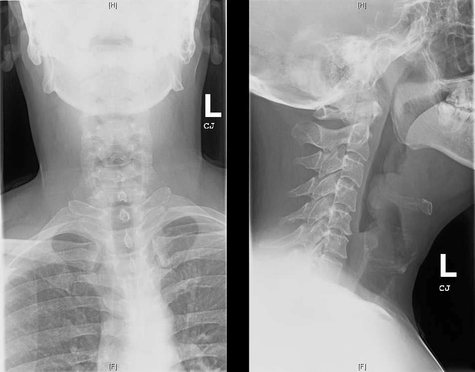
Soft Tissue Neck X-ray:

epiglottitis


THE TALK

What is an epiglottis?

  • Back wall of the vallecular space below base of tongue
  • Infectious epiglottitis = cellulitis of epiglottis
    • Progresses to involve entire supraglottic larynx (including aryepiglottic folds and arytenoids) leading to difficulty breathing

Isn’t epiglottitis a pediatric diagnosis?

  • Incidence decreased in children since haemophilus influenza vaccination
  • Incidence in adults (2006): 1.6 cases per 100,000 adults
    • Usually a/w HTN, DM, Substance abuse or immune deficiency

How do I diagnose it?

  • Fiberoptic nasal laryngoscopy = gold standard
  • Lateral neck x-ray: look for the “thumbprint” sign
  • CT neck (if diagnosis unclear)

What do I do if I suspect/diagnose it?

  • MANAGE AIRWAY- Be prepared for a cricothyrotomy!
  • Have patient sitting up in bed
  • Consider prophylactic intubation
    • Risk of laryngospasm with scope
  • Heliox (mixture of Helium and Oxygen) can be used as a temporizing measure
  • Augmentin or Ampicillin-sulbactam (Unasyn) are preferred initial antibiotics
  • Consider Vancomycin if patient is critically ill and MRSA a possible etiology
  • NSAIDS for pain control
  • +/- Steroids for symptomatic relief

This was too long and I didn’t read it- what should I know?

  • Patient with sore throat and muffled voice but no obvious signs of upper airway obstruction need further work up
  • Have your triple set up ready (direct laryngoscope, video laryngoscope and cricothyroidectomy) and have backup (ENT/Anesthesia) available for a difficult airway

REFERENCES/FURTHER READING:

  • Frantz TD, Rasgon BM, Quesenberry CP. Acute Epiglottitis in Adults: Analysis of 129 Cases. 1994;272(17):1358-1360.
  • Woods, Charles. “Epiglottitis (supraglottitis): Clinical Features and Diagnosis.” UpToDate, 23 June 2015. Web.
  • Rogers, Matt. “Epiglottitis.” Core EM, 26 Aug. 2015. Web.

Aortic Dissection

Follow-up Rounds
1/22/2016
Article inspired by: Dr. Joshua Schwarzbaum, PGY-3


THE CASE

60 y/o F PMHx HTN BIBEMS after bystanders called for erratic behavior. As per triage nurse, patient is rambling while speaking and diaphoretic but with no visible injuries. Patient reports he cannot breathe and that he was told he passed out while walking his dog. He reports he feels cold and sweaty. Denies fevers, chills, nausea, vomiting, chest pain, abdominal pain, or recent travel.

EKG- sinus tachycardia

Physical Exam:
Vitals: BP- 106/74, HR-107, RR-20, Temp-96F, SpO2- 94%, FS- 125
General: Mild distress, lying in bed
Chest: CTA b/l
Cardiovascular- S1/S2, Tachycardic
Abdomen- Soft, NT/ND
Extremities- Warm, No peripheral edema
Neurological- Waxing/waning mental status, PERRL, Moving all extremities, Will not comply with exam

Labs:
138/hemolyzed/108/17.4/14/1.5<265
15.2>15.4/46.3<184
Trop: 0.22, CK: 235

CXR: No acute cardiopulmonary process

CT Head: No acute intracranial pathology

CT Chest: Findings are consistent with bibasilar pulmonary emboli, extremely slow flow through the heart and evidence of heart failure with smooth thickening of the interstitium and geometric polygons formation. Bibasilar atelectasis/infiltrate. Moderate pericardial effusion which is hemorrhagic in nature and with a history of hypertension, rule out dissection of the aorta which is not opacified due to extremely slow flow.

Echocardiogram:


THE TALK

Is there a way to classify aortic dissections?

  • Arbitrary way: Acute (<2 weeks of symptoms) vs Chronic
    • Life threatening complications 2/2 branch involvement or aortic rupture more common in acute
  • Stanford Classification
    • Type A
      • Involving ascending aorta, regardless of site of primary intimal tear
    • Type B
      • Everything else
  • Debakey Classification
    • Type 1
      • Originating in ascending aorta and propagating to at least aortic arch
    • Type 2
      • Originating in and confined to ascending aorta
    • Type 3
      • Originating in the descending aorta and extending distally or proximally

What type of dissection is more common?

  • Ascending aortic dissection ~2x more common than descending aortic dissection
    • Right lateral wall of ascending aorta = most common site
    • Aortic arch involvement occurs in ~30%

How does an aortic dissection happen?

  • Primary event = tear in aortic intima
  • Blood passes into aortic media through tear, creating false lumen
  • Related to shear forces

How can people die from aortic dissection?

  • Rupture of dissection into pericardium leading to cardiac tamponade
  • Acute dissection of aortic valvular annulus leading to severe aortic regurgitation
  • Obstruction of coronary artery ostia leading to MI
  • Abdominal aortic branch vessel obstruction leading to end organ failure

What are some risk factors for aortic dissection?

  • HTN (abrupt, transient, severe increase in BP, ex: crack cocaine or high intensity weight lifting)
  • Collagen disorders (ex: Marfan, Ehler-Danlos, annuloaortic ectasia)
  • Preexisting aortic aneurysm
  • Bicuspid aortic valve (always involves ascending aorta)
  • Aortic instrumentation/cardiac surgery
  • Aortic coarctation
  • Turner syndrome
  • Vasculitis (ex: Giant cell arteritis, Takayasu arteritis, RA, Syphilitic aortitis)
  • Trauma (but usually causes aortic rupture or transection)
  • Pregnancy/Delivery

What are some things I should elicit on my H&P?

  • Acute pain (typically severe, sharp/knife-like)
    • Abdominal pain/Mesenteric ischemia (think celiac or mesenteric arteries involvement)
    • Back or flank pain/Renal failure (think renal artery involvement)
  • Pulse deficit (a weak or absent carotid, brachial, or femoral pulse resulting from the intimal flap or compression by hematoma)
    • Upper extremity pulselessness/Hypotension (think subclavian artery involvement)
    • Lower extremity pain/pulselessness/weakness (think common iliac artery involvement)
  • Heart murmur (new diastolic decrescendo murmur)
    • Aortic insufficiency/Heart failure (think aortic valve involvement)
  • Focal neurologic deficit
    • Stroke/Syncope (think brachiocephalic, common carotid or left subclavian artery involvement)
    • Paraplegia (think intercostal artery involvement as they give off spinal/vertebral artery)
    • Horner syndrome (ptsosis/miosis/anhidrosis) (think superior cervical sympathetic ganglion involvement)
  • Hypotension
    • Cardiac tamponade (think pericardium involvement)
    • MI (think right coronary artery involvement)
    • Hemothorax/Hemoperitoneum (think thoracic or abdominal aorta involvement)

How do I manage someone with aortic dissection?

  • Pain control with narcotics (morphine)
  • If hemodynamically unstable/airway compromise, intubate
  • Heart rate control to <60bpm (IV beta blocker ex: propranolol or labetalol)
    • Labetalol is an alpha- and beta-receptor antagonist (may be more effective in controlling both heart rate and blood pressure as a single agent)
    • Esmolol has short half-life and can be titrated to effect
    • Esmolol also beneficial in asthma or heart failure (pts who are intolerant to BB)
    • Other options if cannot use BB: Verapamil or Diltiazem
  • Reduction of systolic blood pressure of 100-120mmHg
    • If after BB, still elevated- can use Nitroprusside (but only after BB because vasodilation alone induces reflex activation of sympathetic nervous system -> enhanced ventricular contraction -> increased aortic wall shear stress)
    • Can use ACE-inhibitors
    • AVOID HYDRALAZINE (increases aortic wall shear stress)

What if my patient is hypotense?

  • Prior to giving volume, determine cause:
    • Blood loss
    • Hemopericardium with tamponade
    • Valvular dysfunction
    • Left ventricular systolic dysfunction
  • AVOID inotropic agents (increase aortic wall shear stress)

REFERENCES

Schwarzbaum J. “Follow up Rounds: Aortic Dissection” Jacobi Medical Center. Jacobi/Montefiore Emergency Medicine Conference. Bronx. Jan 2016. Case Presentation

Manning, Warren J., and James H. Black. “Clinical Features and Diagnosis of Acute AorticDissection.” Clinical Features and Diagnosis of Acute Aortic Dissection. UpToDate, 19 Feb. 2016. Web.

Manning, Warren J. “Management of Aortic Dissection.” Management of Aortic Dissection. UpToDate, 20 Nov. 2013. Web.

Epidural Abscess

Followup Rounds
12/11/15
Article inspired by: Dr. Anna Meyendorff


THE CASE

27 y/o M p/w left upper back pain x 10 days.

  • Felt a bump on his back for the last few weeks,
  • Swelling waxed and waned
  • Took ibuprofen and has been using a heating pad for the pain.
  • Last 3 days, has developed fevers/chills, nausea, vomiting, diarrhea, and poor appetite.
  • Entire body hurts, especially his muscles
  • Cannot walk secondary to the pain and general weakness
  • Denies numbness, paresthesias, incontinence, IVDU
  • ROS- mild headache with photophobia, some difficulty swallowing and a sensation of food getting stuck in his throat

PMHx: HIV/AIDS (last CD4 6, VL 119,520), Frequent skin abscesses

PSHx: Anal wart resection

Meds: Non-compliant (prescribed: Norvir, Reyataz, Truvada, Azithromycin, Bactrim)

Allergies: Denies

SHx: Smokes 1/2ppd, occasional cocaine, denies IVDU; multiple male partners, uses condoms

Physical Exam:

  • Vitals: BP: 118/50, HR: 116, RR: 20, Temp: 102.6F, SpO2: 97%
  • General: Lying on stretcher in no distress, Cachectic, Diaphoretic, Oral thrush
  • Chest: CTA b/l, No wheezes/rales/rhonchi
  • Cardiac: Tachycardic, No murmurs/rubs/gallops
  • Abdomen: Soft, NT/ND
  • Rectal: Tone intact, No saddle anesthesia
  • Back: No midline cervical/thoracic/lumbosacral tenderness; Large tender area with local induration and calor over the left paraspinal area at mid-thoracic level
  • Neuro: AAOx3; Unable to test gait (refusing 2/2 pain); Motor 5/5 in b/l UE, 5/5 in LLE hip flexion, knee flex/ext, plantar/dorsiflexion, 3/5 RLE hip flexion, 4/5 knee flex/ext, 5/5 plantar/dorsiflexion; Sensation symmetrically intact to light touch over face, trunk, and extremities; Reflexes 3+ b/l biceps, 2+ b/l ankle jerk, toes flex b/l
  • Extremities: Warm and well-perfused x 4, Several scars on right arm (from old-appearing, small, healed abscesses), Tenderness to light touch over R anterior thigh and L triceps

Labs

  • 7.8>10.8/33<322
  • 127/4.7/89/25/19/0.9<107
  • Lactate 1.9, Alb 3.0, T. Bili 1.5, AST 198, ALT 87, Alk Phos 84
  • Coags nl, CRP 391, ESR 117

Imaging

  • CT Chest & Thoracic Spine w/o contrast: Large lobulated collection at the lateral aspect of the left paraspinal musculature concerning for a soft tissue abscess measuring 2 x 6 x 11.7 cm. Small epidural collection along the left lateral aspect of the upper thoracic spine at the T3 level. Possible fluid collection in the neuroforamina at T3/4. No osseous erosions seen. Follow-up with contrast-enhanced MRI recommended.

THE TALK

  • Two types of epidural abscesses
    • Intracranial epidural abscess
    • Spinal epidural abscess (nine times more common)
      • Most common in thoracolumbar areas (larger epidural space and more infection-prone fat tissue)

Why is an epidural abscess dangerous?

  • Can expand and compress brain/spinal cord by
    • Direct compression
    • Thrombosis/thrombophlebitis of nearby veins
    • Interruption of arterial blood supply
    • Bacterial toxins and mediators of inflammation

What does the epidural abscess contain?

  • S. aureus (63%)
  • Mycobacterium tuberculosis (more frequent cause in developing world)
  • In acute cases, frank pus
  • More commonly, granulation tissue (when present > 2 weeks)

How can bacteria get into the epidural space?

  • Hematogenously
    • Skin/soft tissue infections
    • Bacterial endocarditis
    • PNA/UTI
  • Direct extension from infected contiguous tissue
    • Vertebral osteomyelitis
    • Retropharyngeal abscess
    • Psoas abscess
  • Direct inoculation into the spinal canal
    • Epidural injections or catheters
    • Penetrating injury
    • Spinal stimulators

Who is at risk for epidural abscesses?

  • IV drug users
  • Immunocompromised (diabetes, alcoholics, HIV)
  • Recent perispinal procedures (epidural, L.P., spinal surgery)
  • Infection of adjacent structures (vertebral osteomyelitis, infected pressure sores)

Is there a classic triad I should look out for?

  • Fever (50%)
  • Back Pain
  • Neurological deficits (usually absent early on so diagnosis missed at 1st presentation)

What is the classical progression of the disease, if left untreated?

  • Back pain (focal and severe), progressing to
  • Nerve root pain (“shooting” or “electric shocks”), progressing to
  • Motor weakness, sensory changes and bladder/bowel dysfunction, progressing to
  • Paralysis (quickly becomes irreversible)

Are there any lab tests I can order to help me?

  • Leukocyte count can be elevated or normal
  • ESR is usually elevated in both epidural abscess and vertebral osteomyelitis
  • Blood cultures

Hmm, I guess not. How about imaging?

  • MRI is the preferred test (image entire spinal column because multiple skip lesions are common)
    • May require pain management so they can lie flat/still for an MRI
    • MRI is important to distinguish epidural soft tissue edema VS epidural abscess
    • Fluid-equivalent signal intensity on T2-weighted images with rim enhancement and hypointense center
  • CT with IV contrast is an acceptable alternative, if no MRI available

Okay, what do I do if the MRI suggests an epidural abscess?

  • Diagnosis
    • 2 sets of blood cultures
    • Direct needle aspiration (usually under CT guidance) of abscess fluid/pus
    • Do NOT L.P. (risk of seeding subarachnoid space leading to meningitis)
  • Treatment
    • Reduce size and ultimate elimination of inflammatory mass
      • Early surgical decompression and drainage (within 24 hours)
      • Medical (conservative) approach only if lacking risk factors (such as advanced age, bacteremia, WBC > 12,500 cell/L, diabetes, MRSA infection, in whom organism is known from aspiration and no neurological deficit)
    • Eradicate the causative organism
      • Empiric antibiotics against staphylococci, streptococci and gram-negative bacilli
        • Vancomycin (for empiric MRSA) +
        • Nafcillin OR Oxacillin (for optimal MSSA coverage as better than Vanc) +
        • Metronidazole +
        • Cefoxatime OR Ceftriaxone OR Ceftazidime (preferred if Pseudomonas considered)

REFERENCES

Meyendorff A. “Follow up Rounds: Epidural Abscess” Jacobi Medical Center. Jacobi/Montefiore Emergency Medicine Conference. Bronx. Dec 2015. Case Presentation

Sexton, Daniel J., and John H. Sampson. “Spinal Epidural Abscess.” Spinal Epidural Abscess. UpToDate, 4 Jan. 2016. Web.

Nickson, Chris. “Spinal Epidural Abscess.” Spinal Epidural Abscess. Life in the Fast Lane, Web.

Chemical Sedation

C.A.L.M. = Management of Agitated Patients in the Emergency Department
12/9/2015
Article Inspired by: Dr. Vincent Nguyen


THE CASES

  • Young ~70kg male with 2 point restraints in a gurney, attempting to get over the handrails. EtOH level of 346 mg/dL.
  • Middle-aged male brought in by NYPD after barricading himself in an apartment. He is talking nonstop about someone stealing all his belongings.
  • Young make brought in handcuffs yelling racial insults at NYPD with BP noted to be 168/105 mmHg and HR of 136/min.
  • 62 y/o M with known history of EtOH abuse who is standing naked next to his stretcher, tremulous, bleeding from his IV site (that he just removed) and urinating on the ground. BP of 156/92 mmHg and HR of 132/min.

THE TALK

Why would I use medical sedation?

  • Acutely agitated patients pose a danger to the ED staff and to themselves.
  • Even after physical restraints are applied, the patient may still struggle.
  • Medical sedation allows the patient to become calm.

Which medications should I use?

Every physician has their “go-to” sedative, from single agent to combination therapies, using benzodiazepines, antipsychotics, antihistamines and even paralytics!

Here are a few examples when you ask ER docs about their preferred “takedown” medication:

  • Ketamine 0.5-1 mg/kg IV
  • Thiopental + succinylcholine (or rocuronium)
  • Droperidol 5 – 10mg IV
  • Droperidol 5mg IM
  • Droperidol 5mg IM + midazolam 2mg IM
  • Droperidol 5mg IM + midazolam 5mg IM
  • Midazolam 2.5 – 5mg IV
  • Midazolam 10mg IM
  • Haloperidol
  • Haloperidol + midazolam
  • Haloperidol 5mg + lorazepam 4-8mg IV
  • Haloperidol 5mg + lorazepam 2mg + diphenydramine 50mg IV
  • Lorazepam 0.5 – 2mg PO
  • Olanzepine 10mg PO
  • Quetiapine
  • Diazepam 10mg IV
  • Diazepam 10mg IV + fentanyl 100mcg

REMEMBER: “One size does NOT fit all!” Patients are different. Their reason for agitation is different…their level of agitation varies…

Okay, what should I take into consideration when I choose medications?

  • Type and Level of agitation
  • Body size
  • Age
  • Medical history (e.g. drug dependence)
  • Previous response to sedative drugs

What are some of the different types of agitation?

A brief clinical assessment can usually help you categorize the agitation into one of 4 groups:

  • EtOH intoxication
  • EtOH withdrawal
  • Psychosis
  • Stimulant/Undifferentiated

The key is to stay “C.A.L.M.” – it’s a mnemonic that’s easy to remember:

  • Converse with patient
    • Verbally engage the patient (frequently can be successful in <5 min)
    • Establish a collaborative relationship (Offer food/drink or a chair)-
    • Verbally de-escalate out of agitated state
      • Respect personal space
      • Do not be provocative
      • Establish verbal contact
      • Be concise
      • Identify wants and feelings
      • Listen closely to what the patient is saying
      • Agree or agree to disagree
      • Lay down the law and set clear limits
      • Offer choices and optimism
      • Debrief the patient and the staff
  • Activate a team (treat the acutely agitated patient the same way you would a trauma notification)
    • Minimum of 5 team members (preferably ED staff trained in handling behavioral problems)
    • Violence Team Management
    • Overhead announcement (so team members know to go to the designated area)
      • At least 1 team member should be female if the patient is female
    • Team leader should clearly designate roles
    • Team leader signals when to initiate contact
      • DO NOT impair patient’s breathing (i.e. chest, neck or mouth)
      • Explain to patient what is happening at all times
  • Limits and Restraints
    • Supine patient in 4-point restraints on a stretcher
    • One arm up and one arm down
    • Head of bed raised 30 degrees
  • Medications
    • Medications

REMEMBER: An agitated patient doesn’t have to be “put to sleep.” They don’t have to “sleep it off” for 8 hours… Careful choice of medication can simply calm the patient, allowing for a meaningful exam, and disposition in a couple of hours.

What should I monitor after they are sedated?

  • Pulse, RR
  • Pulse Oximetry
  • Blood Pressure
  • Initial temperature should be recorded
  • EKG (if patient is given a QT prolonging agent, especially if administered IV)
  • Close monitoring of airway adequacy
  • Neurovascular observations distal to restraints
  • Remove restraints as soon as possible (start with one leg, then contralateral arm)
  • Debrief patient when calm and explain what happened

TAKE HOME POINT:

Not all agitation is the same so … DON’T treat them all the same!


REFERENCES

Nguyen, V. “Conference: Chemical Sedation” Jacobi Medical Center. Jacobi/Montefiore Emergency Medicine Conference. Bronx. Dec 2015. Lecture

Lulla, Aditya Al, and Manpreet Singh. “The Art of the ED Takedown – FOAM EM RSS.” FOAM EM RSS, 04 Mar. 2015. Web.

Nickson, Chris. “Chemical Restraint.” Life in the Fast Lane, 7 Aug. 2014. Web.

Hyperkalemia

ECG of the Block
11/25/2015
Article Inspired by: Dr. Lauren Williams


THE CASE

CC: ‘I just don’t feel well’

HPI: 

  • 72 yo F PMH ESRD on HD
  • c/o weakness and malaise
  • Near collapse but denies LOC
  • Denies palpitations, SOB, CP, HA, N/V, dysuria, abdominal pain, melena or hematochezia
  • Due for scheduled dialysis on day of presentation

Physical Exam
VSS
General: AAOx3, NAD
HEENT: MMM
CVS: RRR, S1, S2, holosystolic murmur
Pulm: CTAB
Abd: Soft, NT

EKG not done at triage but performed at your request after evaluating patient:

image


THE TALK

Definitions

  • Hyperkalemia = potassium level > 5.5 mEq/L (a/w repolarization abnormalities)
    • Peaked T waves
  • Moderate hyperkalemia = serum potassium > 6.0 mEq/L (a/w progressive paralysis of the atria)
    • P wave widening/flattening
    • PR segment prolongation
    • Eventual disappearance of P waves
  • Severe hyperkalemia = serum potassium > 7.0 mEq/L (a/w conduction abnormalities and bradycardia)
    • Prolonged/bizarre QRS morphology
    • High-grade AV block with slow junctional or ventricular escape rhythms
    • Conduction blocks
    • Sinus bradycardia/slow AF
    • Eventual development of a sine wave appearance
  • Serum potassium level of > 9.0 mEq/L can cause cardiac arrest
    • Asystole
    • Ventricular fibrillation
    • PEA with a bizarre, wide complex rhythm
  • In individual patients, serum potassium level may NOT always correlate with the EKG changes

How do I treat hyperkalemia?

  • First step: Antagonize the cardiac membrane effects of potassium (with calcium)
    • Hypocalcemia can increase the cardiotoxicity of hyperkalemia
    • Effect of calcium begins within a few minutes and lasts for ~30-60 minutes
    • Indicated only when patient has EKG changes
    • Should be administered as infusion given short half-life
  • Second step: Drive the extracellular potassium into the cells (insulin with glucose, sodium bicarbonate, and beta-2 adrenergic agonists)
    • Insulin enhances the activity of the sodium-potassium-ATPase pump in skeletal muscle
      • Effects begins w/in 10-20 minutes, peaks at 30-60 minutes and last for 4-6 hours
      • Glucose is usually given with insulin to prevent hypoglycemia
    • Beta-2-adrenergic agonists (ex: albuterol)- similar to insulin
      • Beta-2-adrenergic receptors in skeletal muscle also activate the inwardly directed Na-K-2Cl cotransporter
      • Additive effect when used with insulin and glucose.
    • Sodium bicarbonate raises serum pH, resulting in hydrogen ion release from the cells -> accompanying potassium movement into the cells
  • Third step: Remove potassium from the body
    • Loop/thiazide diuretics
      •  Limited data to support their use but makes sense clinically to give them if pt not in renal failure
    • Cation exchange resin
      • A/w intestinal necrosis even after one dose (esp in post-op patients, those with ileus, or bowel obstruction)
      • Should only be considered in patients with severe hyperkalemia and ESRD when dialysis is not available
    • Dialysis
  • In a hemodialysis patient, treating hyperkalemia acutely with these medications could drive potassium into the cells, which could then diminish subsequent potassium removal during the dialysis session if performed right afterwards, leading to rebound hyperkalemia after dialysis

TAKE HOME POINT: If a triage ECG is not done for your dialysis patient, that 10 second strip of paper should be at the top of your priority list.


REFERENCES

Williams L. “EKG of the block: Hyperkalemia” Jacobi Medical Center. Jacobi/Montefiore Emergency Medicine Conference. Bronx. November 2015. Case presentation

“ECG Features of Hyperkalaemia.” Burns, Edward. Life in the Fast Lane Medical Blog. LITFL, Web.

“Treatment and Prevention of Hyperkalemia in Adults.” Mount, David. UpToDate, Web.

Drowning

Wednesday Conference
11/11/15
Article Inspired by: Dr. Sui Fai Li and Dr. Andrew Chertoff


THE CASE

20 y/o M BIBEMS after falling into the river. He is pulseless, apneic and cold. He is intubated, and his temperature is 80F. CPR has been in progress for 2 hours. Monitor shows VFib throughout CPR. What do you do?

  • Controversial but keep resuscitating until normothermic (possible neuroprotective effects with hypothermia)

THE TALK

Drowning = “process of experiencing respiratory impairment from submersion/immersion in liquid” as per World Congress on Drowning in 2002

Why is drowning harmful?

  • Initially you can hold breath
  • As some point, due to need for oxygen, will take a deep breath and water will enter airway
  • Clinical picture determined mostly by the amount of water that has been aspirated
    • Water in the alveoli causes surfactant dysfunction and washout
    • Increased permeability of the alveolar-capillary membrane
  • Result = hypoxemia
    • Decreased lung compliance
    • Increased regions of low ventilation to perfusion in the lungs (V/Q mismatch)
    • Atelectasis
    • Bronchospasm

What should I do if I see someone drowning and I am the first person on the scene?

  • Cardiac arrest from drowning is due primarily to lack of oxygen
  • CPR should follow traditional ABC sequence- 5 initial rescue breaths, followed by 30 chest compressions and continuing with 2 rescue breaths and 30 compressions

Should I do the Heimlich maneuver?

  • NO, most frequent complication is vomiting and it delays the initiation of ventilation

How about when the patient is in the Emergency Department?

  • Focus on reversing hypoxemia
    • Non-rebreather mask or nasal cannula if patient is protecting airway
    • If no improvement in 10-30 minutes, intubate
  • If hypothermic, warm them to achieve ROSC and then make a treatment decision
  • ECMO is single most effective way to warm someone, especially if cardiac dysrhythmia
  • CXR in any drowning patient with respiratory systems but initial CXRs usually do not provide prognostic value
  • Be careful about the circumstances (i.e fell off a bridge- now have to worry about trauma related injuries in addition to the drowning)

Is there a way to classify patients?

  • Grade 1 = coughing but normal auscultation
  • Grade 2 = abnormal auscultation; rales in some fields
  • Grade 3 = acute pulmonary edema without arterial hypotension
  • Grade 4 = acute pulmonary edema with arterial hypotension
  • Grade 5 = isolated respiratory arrest
  • Grade 6 = cardiopulmonary arrest

When should I admit a patient?

  • Grade 2 to 6 presentation
  • However, for grade 2, can also place on noninvasive oxygen and if oxygenation normalizes, could discharge after 6-8 hours of observation

Can I send someone home who has had “nonfatal drowning?”

  • Awake and look okay (good arterial oxygenation, no need for supplemental oxygen and no other associated injuries) – check lungs and can be discharged

How should I manage these patients?

  • Neurological
    • Keep head of bed raised
    • Low to normal CO2
    • MAP of 80mmHg
    • Benzodiazepines for seizures
  • Respiratory
    • Protective lung ventilation to prevent ALI and ARDS
    • Bronchodilation
    • ECMO
  • Metabolic
    • Severe metabolic acidosis from lactate
    • Rhabdomyolysis
  • Cardiovascular
    • Extravasation of systemic and pulmonary capillaries + cold diuresis -> hypovolemia
    • SIRS post resuscitation, often require cardiac output monitoring
  • Infection
    • Consider antibiotics if submerged in grossly contaminated water

Terminology

  • Dry drowning
    • Those who do not aspirate liquid into the lungs
  • Wet drowning
    • Aspirate liquid into the lungs
  • Secondary drowning
    • Unrelated event (seizures, cervical spine injury or heart attack) that results in the submersion and subsequent drowning
    • Development of ARDS in recovering victim
  • Freshwater drowning VS Seawater drowning
    • Similar degrees of lung injury, despite difference in osmotic gradients
    • You usually don’t swallow enough water to make any difference to your vascular system
  • Afterdrop Phenomenon
    • When applying external heat to the body, return of pooled, cool blood from previously vasoconstricted extremities -> further lower core temperature = afterdrop
    • Rewarming of trunk first may prevent problem by allowing warm blood to perfuse distally constricted cold extremities
    • More common in dehydrated patients and in patients with frostbitten extremities

REFERENCES

Li, S. “Conference: Drowning” Jacobi Medical Center. Jacobi/Montefiore Emergency Medicine Conference. Bronx. Nov 2015. Lecture

Chertoff, A. “Conference: Drowning Jeopardy” Jacobi Medical Center. Jacobi/Montefiore Emergency Medicine Conference. Bronx. Nov 2015. Lecture

Borghei, Sam, MD, Mizuho Spangler, DO, and Andrew Schmidt, DO. “Drowning Resuscitation.” July 2015. EM:RAP.

Nickson, Chris. “Drowning.” Drowning. LITFL Life in the Fast Lane Medical Blog

Electrical Injuries

Wednesday Conference
11/11/2015
Article Inspired By: Dr. Sui Fai Li and Dr. Andrew Chertoff


-Very little hard data as there are no randomized control trials and mostly just case series

-The first thing to do when someone presents with an electrical injury is to undress the patient and look for any burns/injuries

-Usually, if they present with just numbness, it will resolve

Classification:

  • Low Voltage Injuries
    • Household electrocutions
  • High Voltage Injuries (>1000V)
    • Occupational, Public transit, Transformers

Pathophysiology:

  • Damage done depends on:
    • Amperage (amount)
    • Duration
    • Voltage (force)
    • Resistance
    • and maybe Pathway
  • Amp X Resistance = Volt
    • Higher voltage = higher amperage
      • Household (U.S.) = 120V
      • Public Transit (Subway ~600 to Overhead ~15,000V)
      • Substations ~11,000-400,000V
    • Dry skin 10-40x more resistant than wet skin
    • Alternating current (AC) can be three times worse than Direct current (DC)
      • High-voltage DC is usually a single muscle spasm
      • Alternating current causes prolonged/continuous muscle contraction and tetany leading to longer electrical exposure

What type of injuries can you see with electricity?

  • Trauma
  • Thermal injury
  • Electrical injury
  • Burns
    • Arc burns
    • Crease burns

What organ systems do I have to worry about?

  • Neurological
    • LOC, transient paralysis, or paresthesias
    • Keraunoparalysis (fixed, dilated pupils; lower extremity cyanosis; paralysis from vasospasm)
      • Classically with high voltage lightning injuries
      • Continue resuscitation for prolonged period of time despite fixed, dilated pupils
    • Most permanent injuries are secondary events
  •  Cardiac
    • A/C induces ventricular fibrillation whereas D/C induces asystole
      • Lightening causes massive direct countershock which depolarizes entire myocardium causing asystole
    • Cardiac contusion common
    • Acute MI is rare
  • Vascular
    • Thrombosis/hemorrhage
    • Labial artery
      • Usually 3rd degree, 6-20% bleed, need referral to specialty
  • Ophthalmology
    • Lightning strike associated cataracts (need good follow-up)
  • Respiratory- Aspiration
    • Lightning can induce paralysis of medullary center leading to primary respiratory arrest
  • Musculoskeletal
    • Rhabdomyolysis
    • Compartment syndrome
  • Obstetrics
    • Stillbirths
    • Lightning strikes – fetal mortality close to 50%
      • First trimester- confirm fetal heart tone, d/c with spontaneous abortion precautions
      • Second/Third trimester- OB for fetal monitoring and significant risk for placental abruption

Okay, what should I do in the trauma bay?

  • Airway
    • May require intubation if intubated, major burns or for specific treatment (suxamethonium safe for 48 hours)
  • Breathing
    • Lung protective ventilation strategy
    • Look for same life threatening chest injuries as in trauma patients
  • Circulation
    • Prevent effects of rhabdomyolysis (hyperkalemia, hypocalcemia, hyperphophatemia, metabolic acidosis)
    • Restore normal circulating volume
      • May require ionotropes/vasopressors if SIRS
    • Parkland’s formula
  • Disability
    • R/o associated TBI
    • Prevent secondary brain injury
    • Normoglycemia
  • Exposure
    • Quantify severity of burns (depth, percent total body surface area)

Who should I admit and who can I send home?

  • Low voltage, asymptomatic- Do nothing and discharge
  • Low voltage, mild symptoms- Monitor and can discharge, if EKG and UA normal
  • High voltage (>1000V)- Admit for monitoring

REFERENCES:

Li, S. “Conference: Electrical injuries” Jacobi Medical Center. Jacobi/Montefiore Emergency Medicine Conference. Bronx. Nov 2015. Lecture

Chertoff, A. “Conference: Electrical injuries Jeopardy” Jacobi Medical Center. Jacobi/Montefiore Emergency Medicine Conference. Bronx. Nov 2015. Lecture

“Episode 1.0 – Electrical and Lightning Injuries.” Core EM

Nickson, Chris. “Electrical Injury.” Electrical Injury. LITFL Life in the Fast Lane Medical Blog


Thyroid Storm

Follow-Up Rounds (10/2/15)
Article Inspired by: Adelle Iusim, PGY-3


THE CASE:

CC: Body aches, Fever, Left-sided abdominal pain x 3 weeks

Triage Vitals- BP-125/78, HR: 99, RR: 18, SpO2-93%, Temperature: 100.8F

HPI: 25 y/o F presents with abdominal pain (left sided, generalized cramping), subjective fever/chills, and myalgias x 3 days. She reports persistent nausea and vomiting with PO intake. She also endorses urinary frequency and a foul smelling urine.

PMHx: Hyperthyroidism, Seizures(?), Latent tuberculosis
PSHx: none
Meds: cannot recall
Allergies: NKDA

Pertinent positives on physical exam:
HEENT: Dry mucous membranes
Cardiovascular: Tachycardic
Abdomen: BS+ x4, Soft, NT/ND
Back: B/l CVA tenderness

EKG: Sinus tachycardia @130bpm

Differential Diagnosis:

  • Infectious:
    • Influenza
    • Viral Syndrome
    • Strep
    • Pneumonia
    • Encephalitis
    • Meningitis
    • Intraabdominal pathology
    • UTI
    • Pyelonephritis
  • Cardiovascular/Endocrine:
    • Arrythmia
    • Hyperthyroidism
    • Thyroid Strom
    • Toxidrome due to drugs
    • Pheochromocytoma

Labs:
Na: 135, K: 4.7, Cl: 102, HCO3: 20.8, BUN: 15, Cr: 0.4, Glu: 97

Ca: 9.3, Mg: 2.2,T. Prot: 8.3, Albumin: 4.1, T. Bili: 1.2, Alk Phos: 232, AST: 25, ALT: 35; AG: 12.2

WBC: 13.5, Hgb: 13.8, Hct: 41.1, Plt: 231, MCV: 81, Gr: 66.9, Lym: 26.2

UA: Cloudy, glucose negative, bili negative, ketones trace, sp grav: 1.02, large blood; +nitrite; large leuk esterase; WBC: 479; RBC: 191; Bacteria 4+; Epithelial 81; Micro: WBC: many, RBC: 7-10, Bacteria: many

CXR- no acute cardiopulmonary process

TSH: <0.008, Free T4: 11.13

Management:

After further questioning (and eliciting palpitations, weight loss, insomnia on ROS) and further examination (significant for mild exopthalmos and hyperreflexia), she was given Tylenol, 1L NS, Ceftriaxone 1g IV and endocrine was called for likely Grave’s disease.

Endocrine consult: Methimazole 30mg orally; Wait 1 hr, give 5 drops of 0.25mL Potassium Iodide sski q6hr; Atenolol 50mg q8hr (with titration to HR of 80); Check TPO and TSI antibody; Consider steroids if patient deteriorates.

Hospital Course:

Continued on Atenolol, Methimazole. Hydrocortisone 100mg q8hr was started, 0.25mL SSKI potassium iodide q6hr was restarted.

Continued treatment with Ceftriaxone for 2 days, and once cultures came back as pansensitive, switched to Ciprofloxacin for 2 days.

TSI 5.9 (<1.3 is normal), Thyroid peroxidase antibody <10 (<35 is normal)

Discharged after 2 days on Atenolol 50mg TID and Methimazole 30mg daily with endocrine outpatient followup.


THE TALK

Thyroid Storm:

  • Rare, life threatening exacerbation of hyperthyroid state with 1 or more organ dysfunction
  • Unclear etiology-
    • Rapid rate of increase in serum thyroid hormone levels
    • Increased responsiveness to catecholamines
    • Enhanced cellular responses to thyroid hormone

WHEN DOES THYROID STORM OCCUR?

  • Systemic insult
    • Infection
    • Trauma
    • Surgery (usually 6-24 hours post)
    • Hyperosmolar coma
  • Endocrinal insult
    • DKA
  • Drug/Hormone-related
    • Withdrawal of thyroid medication
    • Acute iodine load
    • Thyroid gland palpation
    • Ingestion of thyroid hormone
  • Cardiovascular insult
    • MI
    • CVA
    • PE
  • Pregnancy related
    • Parturition
    • Eclampsia

WHAT EFFECTS CAN IT HAVE ON THE BODY?

  • Direct ionotropic and chronotropic effects:
    • Decreased systemic vascular resistance
    • Increased blood volume
    • Increased contractility
    • Increased CO
  • Exaggeration of hyperthyroidism symptoms:
    • Fever (104F-106F)
    • Tachycardia >140 beats/min
    • Altered Mental Status (agitation, anxiety, delirium, psychosis, stupor, coma)
    • Severe nausea, vomiting, diarrhea, abdominal pain, hepatic failure with jaundice
    • Congestive heart failure
    • Cardiac arrhythmia (severe tachycardia or atrial fibrillation in 10-35% cases)
    • Enhanced contractility → elevations in systolic BP and pulse pressure –> dicrotic or water-hammer pulse
    • Death

WHAT SHOULD I LOOK FOR IN MY PHYSICAL EXAM?

  • Goiter
  • Exopthalmous (to think about Grave’s)
  • Lid lag
  • Hand tremor
  • Warm and moist skin

WHAT OTHER DIAGNOSES SHOULD I BE CONSIDERING?

  • Infection/Sepsis
  • Sympathomimetic ingestion (ex: cocaine, amphetamine, ketamine drug use)
  • Heat exhaustion
  • Heat stroke
  • Delirium tremens
  • Malignant hyperthermia
  • Malignant neuroleptic syndrome
  • Hypothalamic stroke
  • Pheochromocytoma
  • Medication withdrawal (ex: cocaine, opioids, etc)
  • Psychosis
  • Organophosphate poisoning

WHAT LAB TESTS SHOULD I ORDER?

  • Chemistry
    • Creatinine may be low
    • Mild hypercalcemia (hemoconcerntration and enhanced bone resorption)
    • Mild hyperglycemia (Catecholamine induced inhibition of insulin release and increased glycogenolysis)
  • CBC
    • Thrombocytopenia
    • Leukocytosis or Leukopenia
  • TFTs (degree of thyroid hormone excess not more profound than uncomplicated thyrotoxicosis)
    • Low TSH
    • High free T4 and or T3
  • LFTs
  • Cortisol level (to rule out concurrent adrenal insufficiency)

IS THERE A CLINICAL TOOL I CAN USE TO DIAGNOSE IT?

Burch and Wartofsky scoring system (sensitive but not very specific)

  • Thermoregulatory Dysfunction
    • Temp 99 to 99.9F = 5 points
    • Temp 100 to 100.9F = 10 points
    • Temp 101 to 101.9F = 15 points
    • Temp 102 to 102.9F = 20 points
    • Temp 103 to 103.9F = 25 points
    • Temp > 104F = 30 points
  • CNS effects
    • Mild (agitation) = 10 points
    • Moderate (delirium, psychosis, extreme lethargy) = 20 points
    • Severe (seizure, coma) = 30 points
  • GI-Hepatic dysfunction
    • Moderate (abdominal pain, nausea, vomiting, diarrhea) = 10 points
    • Severe (unexplained jaundice) = 20 points
  • Cardiovascular dysfunction
    • Tachycardia
      • HR 99-109 = 5 points
      • HR 110-119 = 10 points
      • HR 120-129 = 15 points
      • HR 130-139 = 20 points
      • HR >140 = 25 points
    • Atrial fibrillation = 10 points
  • Heart Failure
    • Mild (pedal edema) = 5 points
    • Moderate (bibasilar rales) = 10 points
    • Severe (pulmonary edema) = 15 points
  • Precipitant history
    • Negative = 0 points
    • Positive = 10 points
  • If score >45: highly suggestive of thyroid storm
  • If score 25-44: impending storm
  • If score <25: thyroid storm unlikely

HOW DO I MANAGE THIS?

  • Supportive care
    • IVF with dextrose (to replenish glycogen stores)
    • MVI/Thiamine (to prevent Wernicke’s when giving dextrose)
    • Tylenol (not NSAIDS or ASA)
      • Aspirin can increase serum free T4 and T3 by interfering with protein binding
    • Bile acid sequestrants (ex: Cholestyramine- to decrease enterohepatic recycling of thyroid hormones)
  • Block peripheral effect of thyroid hormone
    • Beta blocker (to control symptoms/signs induced by increased adrenergic tone- slows HR, increases diastolic filling and decreases tremor)
  • Stop the production of thyroid hormone
    • Thionamide: Methimazole or PTU (slow conversion of T4 to T3 in periphery)
      • PTU favored over Methimazole acutely because of PTU’s effect to decrease T4 to T3 conversion
      • Methimazole has a longer duration of action, is less hepatotoxic, and over weeks of treatment, may result in more rapid normalized of serum T3 than PTU
    • Glucocorticoids (to reduce T4 to T3 conversion, promote vasomotor stability and possible treat associated relative adrenal insufficiency)
    • Iodinated radiocontrast agent (to inhibit peripheral conversion of T4 to T3)- not available in US
  • Inhibit hormone release
    • Iodine solution 1-2 hours after (to decreases release of thyroid hormone from thyroid)
      • Delayed for an hour to prevent iodine from being used as substrate for new hormone synthesis

WHAT IF THEIR SCORE IS 25-44 (IMPENDING STORM)?

  • Beta blocker (ex: propranolol 60 to 80 mg q4-6hr until HR controlled)- can require high doses because of increased drug metabolism as a result of hyperthyroidism
    • Can alternatively use Esmolol (loading dose 250-500mcg/kg, followed by infusion at 50-100mcg/kg per minute)
    • If beta blockers contraindicated, can use CCB (ex: Diltiazem)
  • Propylthiouracil 200mg q4hr OR Methimazole 20mg q4-6hr
  • 1 hour after first dose thionamide taken, Iodine (saturated solution of potassium iodide [SSKI]) 5 drops q6hr OR Lugol’s solution 10 drops q8hr

AND IF THEIR SCORE IS > 45 (LIKELY THYROID STORM)?

In addition to the above,

  • Glucocorticoids (Hydrocortisone 100mg IV q8hr)
  • Cholestyramine 4g QID
  • Treatment of any precipitating factors
  • Some patients require fluids while others require diuresis (ex: CHF)
  • Tylenol instead of ASA

IS SURGERY EVER AN OPTION?

  • Surgery is treatment of choice for patients who cannot take thionamide (ex: due to agranulocytosis or hepatotoxicity or allergy)
  • Still require preoperative treatment with:
    • Beta blockers (propranolol)
    • Glucocorticoids
    • Bile acid sequestrants (cholysteramine 4mg orally QID)
    • Patients with Grave’s disease: Iodide (SSKI) 5 drops q6hr or Lugol’s 10 drops q8hr)
    • If not effective, plasmapheresis (removes cytokines, antibodies and thyroid hormones from plasma)

REFERENCES

Iusim, A. “Follow up Rounds: Thyroid Storm” Jacobi Medical Center. Jacobi/Montefiore Emergency Medicine Conference. Bronx. Oct 2015. Case Presentation

Ross, Douglas S., MD. “Thyroid Storm.” UpToDate, 12 Feb. 2015. Web.

Tintinalli, Judith E., and J. Stephan. Stapczynski. Tintinalli’s Emergency Medicine: A Comprehensive Study Guide. 7th ed. New York: McGraw-Hill, 2011.

Weingart, Scott. “Podcast 149 – Thyroid Storm.” Podcast 149- Thyroid Storm. Emcrit, 16 May 2015. Web. <http://emcrit.org/podcasts/thyroid-storm/&gt;.

Massive Transfusion Protocol

Trauma Rounds (9/23/15)

Article Inspired by: Dr. Paul Mello (EM PGY-4) and Dr. Nikita Shah (Surgery PGY-4)


THE CASE

10:06am: EMS Notification- 89 y/o M, pedestrian struck, No LOC, alert and oriented, Takes Plavix, Hypotensive on scene = level 1 trauma notification

Initial ER Vitals:

BP: 80/palpable, HR-75, RR-18, Temp-unable to obtain, SpO2-89% (room air)

Primary Survey:

  • A: Airway intact, phonating, no trauma noted
  • B: B/l breath sounds, Equal chest rise
  • C: Hypotensive, Palpable distal pulses, Right 18g antecubital IV noted via EMS, Left 18g antecubital IV placed
  • D: Initial GCS 15, PERRL (3mm b/l), Able to move all fingers/toes, Answering questions appropriately
  • E: Log rolled; No palpable step-offs, mid thoracic spine tender to palpation; Normal rectal tone, no gross blood; Left lower leg deformity with tibia protruding through midshaft anterior/medial aspect

AMPLE History:

  • A: No known medical allergies
  • M: Plavix, ASA
  • P: DM, CAD (s/p stents), Diverticulitis (s/p operative intervention), Malaria
  • L: cannot recall last meal
  • E: Reports struck by vehicle while crossing street and denies LOC

Resuscitation:

  • FAST negative
  • Intubated for airway protection with video laryngoscopy using ketamine and succinylcholine
  • R femoral cordis placed and started on MASSIVE TRANSFUSION PROTOCOL– received 4 units PRBCs, 4 units FFP, and 1 unit platelets
  • Fractured leg splinted
  • Started on Levophed and taken for additional imaging
  • CT head: small subarachnoid/sub-pial hemorrhage
  • CT chest/abdomen/pelvis: Moderate b/l pleural effusions, L PTX 30-40%, Small area of hemorrhage w/in R pleura, L 2nd rib displaced/comminuted fractures, Left 3-10 displaced rib fractures, Comminuted fracture of L scapula
  • CT cervical/thoracic/lumbar spine: Fracture through superior aspect Fracture through superior aspect of T8 vertebral body with displacement of anterior cortex and hematoma, Minimally displaced fracture of T7 tranverse process, Chronic depression deformity of L1 vertebral body
  • Bilateral chest tubes placed with 1200cc of blood noted to drain from R chest tube
  • Admitted to Surgical ICU


THE TALK

MASSIVE TRANSFUSION PROTOCOL

  • Used in trauma, major surgeries, GI bleeds, OB hemorrhage, etc and a/w high mortality
  • Various definitions
    • Replacing patient’s total blood volume in <24 hours
    • Acute administration of >1/2 patient’s blood volume an hour
  • Physiologically, compensatory mechanisms can maintain perfusion to vital organs until ~30% total body volume loss

WHY HAVE A PROTOCOL INSTEAD OF JUST MASSIVE TRANSFUSION?

  • To stop the lethal triad of coagulopathy, acidosis, and hypothermia
  • Coagulopathy
    • Tissue trauma → activation and consumption of coagulation factors
    • Prolonged shock, Hypoxia, Hypothermia, Failure to clear activation peptides (act as competitive inhibitors) → Reduced activity of coagulation factors
    • Blood replacement → Dilutional effects on coagulation proteins and platelets
  • Acidosis (excess protons)
    • Interferes with assembly of coagulation factor complexes involving calcium and negatively-charged phospholipids → delayed production and reduced concentrations of generated thrombin → delayed fibrin production, altered fibrin structure, and increased susceptibility to fibrinolysis
  • Hypothermia
    • Reduces the enzymatic activity of plasma coagulation proteins
    • Prevents activation of platelets via traction on the glycoprotein Ib/IX/V complex by von Willebrand factor
      • Seen at core temperatures of 34ºC and below

HOW DO I KNOW WHAT TO REPLACE?

  • Crystalloid solutions will replace the deficit in blood volume
  • Red cells will improve and maintain tissue oxygenation
  • HOWEVER, both of these will result in gradual dilution of plasma clotting proteins
    • Adults: ~10% decrease in concentration of clotting proteins for each 500mL blood loss replaced
    • If coagulation protein levels falls <25% of normal (usually after receiving 8-10units red cells in an adult), additional bleeding can occur
    • Therefore, should monitor PT, aPTT, fibrinogen in massive transfusion protocol
    • If PT, aPTT, fibrinogen > 1.5 times the control, 2-8 units of fresh frozen plasma
      • Each unit of fresh frozen plasma increases clotting protein levels by ~2.5%
      • Cryoprecipitate can be used when fibrinogen levels <100mg/dL
  • Both of these will also have similar dilutional effect on platelet concentration
    • Adult: ~10-12 units of transfused RBCs are associated with a 50% fall in platelet count
      • Each unit of platelet should increase platelet count by 5000/microL
  • THEREFORE, should measure PT, aPTT, fibrinogen, platelet count after ~5-7 units of red cells

OKAY, BUT WHAT ABOUT WHEN I AM IN THE TRAUMA BAY?

  • For trauma, hemodynamic stability is the key indicator for transfusion:
    • Expected/ongoing bleeding
    • Dropping hemoglobin
    • Hemorrhagic shock/low systolic blood pressure/tachycardia
  • In trauma, goal is 1:1:1 (units of transfused FFP to units of platelets to units of pRBCs)
    • Blood transfused will have
      • Coagulation factor concentration of 65% of normal
      • Platelet count of 88 x 109/L
      • Hematocrit of 29%
    • BUT taking into account that 30% of platelets and 10% of RBC administered will not circulate, effective concentrations of blood given:
      • Plasma coagulation factor concentration of 65%,
      • Platelet count of 55 x 109/L
      • Hematocrit of 26%

HMM, WHAT CAN GO WRONG?

  • Inadequate resuscitation
    • Administering excessive crystalloids (if patient presenting with uncontrollable hemorrhage/shock, typically have lost 30-40% of blood volume)
      • >50% dilution of coagulation factors
      • Diminution of thrombin generation
    • Hypoperfusion
      • Lactic acidosis
      • SIRS
      • Disseminated intravascular coagulation
      • Multiorgan dysfunction
  • Over-resuscitation
    • High arterial/venous pressures → can dislodge hemostatic clots
    • Increased hydrostatic pressure → interstitial edema → abdominal compartment syndrome
    • Transfusion associated circulatory overload (usually elderly, small children and patients with compromised left ventricular function)
  • Transfusion related issues
    • Metabolic alkalosis
      • pH of unit of blood is 7.10 at 37C due to citric acid
      • pH drops 0.1 pH unit/week due to production of lactic and pyruvic acids by the red cells
      • Metabolism of citrate in each unit of blood → 23 meq of bicarbonate
      • With renal ischemia, excess bicarbonate cannot be excreted in the urine → metabolic alkalosis
    • Hypokalemia
      • Potassium moves into cells in exchange for hydrogen ions that move out of the cells to minimize the degree of extracellular alkalosis
    • Hypocalcemia
      • Each unit of blood anticoagulated with ~3g citrate
      • Healthy adult liver metabolizes 3g citrate every 5 minutes
      • Transfusion at rates higher than 1 unit every 5 minutes or impaired liver function can lead to higher levels of citrate (which binds to ionized calcium)
    • Hypothermia
      • 6 units of RBCs at 4C will reduce body temperature of 70kg adult by 1C
      • Evaporative heat loss from open body cavity (ex: surgery)- decrease by 1C in 40 minutes
    • Hyperkalemia (in infants or patients with renal impairment)
      • Potassium leakage due to prolonged blood storage or irradiation
    • ARDS/TRALI (Transfusion related acute lung injury)

REFERENCES

Melo, P., Shah, N. “Trauma Rounds: Massive Transfusion Protocol” Jacobi Medical Center. Jacobi/Montefiore Emergency Medicine Conference. Bronx. Sep 2015. Lecture

Hess, John R., MD, MPH. “Massive Blood Transfusion.” Massive Blood Transfusion. UpToDate, Feb. 2015. <http://www.uptodate.com/contents/massive-blood-transfusion?source=search_result&search=massive%2Btransfusion%2Bprotocol&selectedTitle=1~150&gt;

“Transfusion for Massive Blood Loss.” Transfusion for Massive Blood Loss. Trauma.org. <http://www.trauma.org/archive/resus/massive.html&gt;.

Patil, Vijaya, and Madhavi Shetmahajan. “Massive Transfusion and Massive Transfusion Protocol.” Indian Journal of Anaesthesia. Medknow Publications & Media Pvt Ltd, Sept-Oct. 2014. <http://www.ncbi.nlm.nih.gov/pmc/articles/PMC4260305/&gt;.