Author: Dr. Neil McCormack, PGY-4
Sometimes it IS a spider bite
Trigger warning: Pictures of spiders (But, seriously, did you expect something different?)
Cutaneous skin abscesses are a common complaint in emergency rooms and have become more prevalent as the years continue.1 According to bacteriology studies, the majority of these abscesses are caused by skin flora, primarily Staphylococcus and Streptococcus species.2, 3 If you ask the patients, however, abscesses are caused by spider bites approximately 100% of the time. Is this reputation warranted? Are spiders the bane of Emergency Medicine’s existence and the source of all of our abscesses all these years?
Information on the clinical presentation of spider bites continues to be unreliable, being based on case reports, case series and databases. For instance, the Bureau of labor 2014 statistics reported 1,470 incidents involving spider bites. The listed symptoms include 230 incidents of “cuts or lacerations,” 300 incidents of “soreness and pain,” and 900 “other” incidents. The data, however, represents only spider bites reported from the workplace, and lack expert confirmation of the actual spider involved.
Before we discuss the various venomous spiders, let’s define the terms. Many individuals use the terms “poisonous” and “venomous” interchangeably but they’re not exactly the same. Poisons are typically secreted by a plant/animal (think poison dart frogs of the tropics) and are introduced into the body by ingestion or absorption through the skin. Venom, by contrast, is a substance that is actively injected into the body by the animal in question.
There is controversy regarding the number of venomous spiders in the United States. Some sources list only the black widow and the brown recluse spiders; other sources will also include the hobo spider. The CDC, for example, only lists two species; however, we will discuss the three classically considered venomous spiders.4
Black Widow
The black widow spider (genus: Latrodectus) is probably the most recognized spider on this list. There are three subspecies in the US named for their primary residence. The Northern black widow (spp variolus) spans primarily northeast from Canada to Florida and as far West as Texas. The Southern black widow (spp mactans) is found throughout the southeastern states. The Western black widow (spp hesperus) is found from Canada to Mexico along the West coast and as far East as Texas.

Black Widow Spider
The black widow spider grows to approximately 0.5 inches in body length as an adult. They can be identified by their distinctive black body with red hourglass marking.
Among black widow spiders, the larger female is the one who can bite and cause symptoms — males and juveniles have fangs that are too small to cause harm to humans. The black widow injects latrotoxins that attack nerves leading to episodic release of neurotransmitters acetylcholine, norepinephrine and GABA.5 This leads to a constellation of symptoms known as latrodectism. Symptoms start at the site of the bite (a pair of red spots, although the bite is commonly unnoticed), then spread contiguously. They initially include severe pain starting within 5-10 minutes of the bite and increasing in intensity over the first hour. Secondarily, generalized muscle pain, abdominal cramps, nausea and vomiting are seen. Piloerection and profuse diaphoresis at the site are also common. Some victims will see symptoms spread to more diffuse pain, muscle cramping, and diaphoresis/piloerection distant from the bite site, though this is rare. The symptoms in pregnant women can sometimes be severe enough to be confused with contractions.6
Treatment involves symptomatic management of pain and cramping with analgesics and muscle relaxants. For severe symptoms, there is an antivenin available,7,8 though the antivenin is generally limited to envenomation treatment center.9,10,11 Symptoms (and here is the part you don’t want to hear) typically resolve in several days but some people can have lingering symptoms for several weeks.12
Brown Recluse
The brown recluse (Loxosceles reclusa) is a spider native to the Southern US. Their distribution lies along the Southern states from Texas to the East coast and as far North as the Ohio valley. How are you, a well-mannered “keep to yourself” kind of person likely to encounter a brown recluse if you live outside of this spider’s habitat? Unfortunately for you, there are reports of brown recluse spiders being transported to other areas of the country via shipping of goods throughout the US. The reports are of singular incidents and not always verified as brown recluse spiders. But it is something to consider, albeit it is exceedingly rare if you do not live in recluse country. This species has a reputation for being dangerous though, like most spiders, they are rarely aggressive towards humans and many bites are defensive.

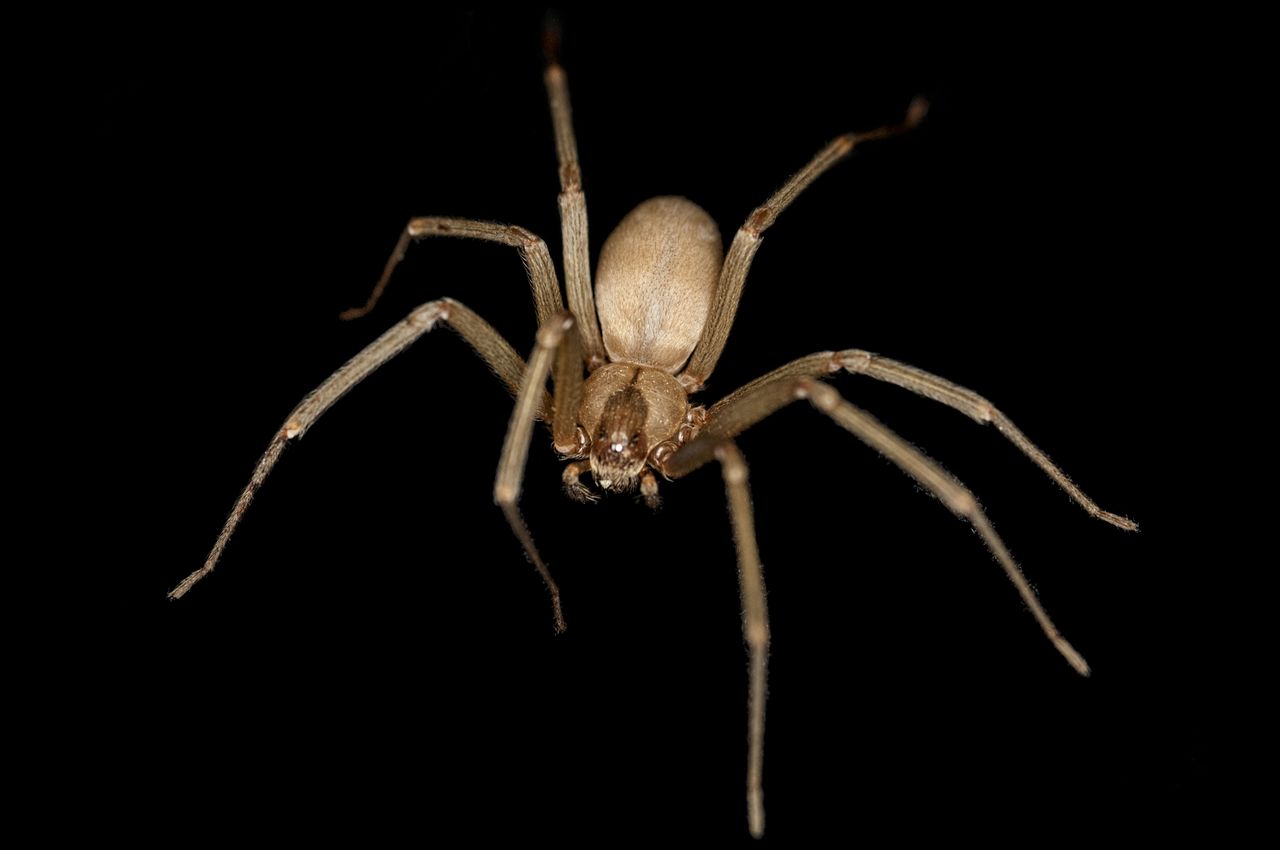
Brown Recluse Spider
The brown recluse spider is, as the name suggests, a brown colored spider that can grow to upwards of 0.75 inches. Their shade can range from dark grey/brown to the lighter color seen in the figure. Luckily for us, the brown recluse has short fangs and are not overly powerful. Thus, most bites require direct skin contact without clothing interference.
The bite of a brown recluse is typically painless initially. The dangerous aspect of this species is that they use a hemotoxin as their venom. This can cause local necrosis of the skin (loxoscelism) due to the toxic protein actions. These main constituents of the venom are hyaluronidase, sphingomyelinase-D, alkaline phosphatase, esterase, and ATPase.13 The clinical manifestations can range from a localized urticarial response only, to forming a blistering, bleeding, ulcerating lesion. The lesion may increase in size and the central blister may become necrotic over the next few days.14 Systemic symptoms, which are rare in the US, include nausea, vomiting, rashes, fevers and joint pains.14 Once necrosis has set in, what do you do now? The most prudent management of these lesions starts with wound care, immobilization, tetanus prophylaxis, and analgesics. Local tissue damage and destruction may be surgically debrided though this will leave a large wound (which may need grafting and prolonged healing) or allowing the wound to for an eschar then heal by secondary intention. One thing to remember when dealing with suspected recluse bites outside of their natural habitat is that if the spider was not caught red-handed there are numerous other causes of skin lesions/necrosis in the medical literature.
Hobo Spider
The hobo spider (Eratigena agrestis) is known as the “aggressive house spider.” I will include this spider here but here is a disclaimer: There is currently debate on whether or not this spider is truly venomous or not. That being said, the hobo spider is native to Europe but in the US it resides in the upper Northwestern US from Canada to Northern California and West towards the Rocky Mountains.

Hobo Spider
The hobo spider is a small brown spider mostly living in fields and outdoors. They grow to approximately 0.5-0.75 inches in length.
Are their bites venomous to humans though? The classic hobo spider bite is described as a small (several cm) painful bite with central erythema leading to blistering within 24 hours. This blistering then often times ruptures leaving a small necrotic lesion which typically will heal within 1-2 months. Only supportive treatment is available for these situations.
There is controversy regarding whether or not this truly happens with hobo spider bites or if this is misattributed to other causes. Some researchers suggest that the bites are caused by MRSA.15 One author reports that out of 33 confirmed Hobo Spider bites, none showed dermatonecrosis.16 The possibility of misattribution of Hobo bites to Recluse spiders or other causes of dermonecrosis leaves us, as well as other researchers,8,15,17,18,19,20 questioning the true nature of hobo spider bites and their medical importance. At this time, if you live in the Northwestern US, let’s just say older reports implicate the hobo spider though this may be an unlikely cause of your patient’s necrosis and should be taken with a certain degree of skepticism.
References
- Taira, B.R., Singer, A.J., Thode Jr, H.C., and Lee, C.C. (2009). National epidemiology of cutaneous abscesses: 1996 to 2005. The American Journal of Emergency Medicine 27, 289–292.
- MEISLIN, H.W., LERNER, S.A., GRAVES, M.H., McGEHEE, M.D., KOCKA, F.E., MORELLO, J.A., and ROSEN, P. (1977). Cutaneous AbscessesAnaerobic and Aerobic Bacteriology and Outpatient Management. Ann Intern Med 87, 145–149.
- Summanen, P.H., Talan, D.A., Strong, C., McTeague, M., Bennion, R., Thompson, J.E., Väisänen, M.-L., Moran, G., Winer, M., and Finegold, S.M. (1995). Bacteriology of Skin and Soft-Tissue Infections: Comparison of Infections in Intravenous Drug Users and Individuals with No History of Intravenous Drug Use. Clin Infect Dis. 20, S279–S282.
- CDC – Venomous Spiders – NIOSH Workplace Safety and Health Topic.
- Ushkaryov, Y.A., Rohou, A., and Sugita, S. (2008). α-Latrotoxin and Its Receptors. Handb Exp Pharmacol 171–206.
- Wolfe, M.D., Myers, O., Caravati, E.M., Rayburn, W.F., and Seifert, S.A. (2011). Black widow spider envenomation in pregnancy. J. Matern. Fetal. Neonatal. Med. 24, 122–126 (2005).
- Singletary, E.M., Rochman, A.S., Bodmer, J.C.A., and Holstege, C.P. (2005). Envenomations. Med. Clin. North Am. 89, 1195–1224.
- Weinstein, S., Dart, R., Staples, A., and White, J. (2009). Envenomations: an overview of clinical toxinology for the primary care physician. Am Fam Physician 80, 793–802.
- Heard, K., O’Malley, G.F., and Dart, R.C. (1999). Antivenom therapy in the Americas. Drugs 58, 5–15.
- Kunkel, D.B. (1984). Arthropod envenomations. Emerg. Med. Clin. North Am. 2, 579–586.
- Quan, D. (2012). North American poisonous bites and stings. Crit Care Clin 28, 633–659.
- Peterson, M.E. (2006). Black widow spider envenomation. Clin Tech Small Anim Pract 21, 187–190.
- Swanson, D.L., and Vetter, R.S. (2006). Loxoscelism. Clinics in Dermatology 24, 213–221.
- Wasserman, G.S., and Anderson, P.C. (1983). Loxoscelism and Necrotic Arachnidism. Journal of Toxicology: Clinical Toxicology 21, 451–472.
- Gaver-Wainwright, M.M., Zack, R.S., Foradori, M.J., and Lavine, L.C. (2011). Misdiagnosis of spider bites: bacterial associates, mechanical pathogen transfer, and hemolytic potential of venom from the hobo spider, Tegenaria agrestis (Araneae: Agelenidae). J. Med. Entomol. 48, 382–388.
- McKeown, N., Vetter, R.S., and Hendrickson, R.G. (2014). Verified spider bites in Oregon (USA) with the intent to assess hobo spider venom toxicity. Toxicon 84, 51–55.
- Bennett, R.G., and Vetter, R.S. (2004). An approach to spider bites. Erroneous attribution of dermonecrotic lesions to brown recluse or hobo spider bites in Canada. Can Fam Physician 50, 1098–1101.
- Fisher, R.G., Kelly, P., Krober, M.S., Weir, M.R., and Jones, R. (1994). Necrotic arachnidism. West J Med 160, 570–572.
- Vetter, R. (2008). Medical aspects of spider bites. Annual Review of Entomology 53, 409–429.
- Vetter, R.S., Roe, A.H., Bennett, R.G., Baird, C.R., Royce, L.A., Lanier, W.T., Antonelli, A.L., and Cushing, P.E. (2003). Distribution of the medically-implicated hobo spider (Araneae: Agelenidae) and a benign congener, Tegenaria duellica, in the United States and Canada. J. Med. Entomol. 40, 159–164.