Hyperkalemia

ECG of the Block
11/25/2015
Article Inspired by: Dr. Lauren Williams


THE CASE

CC: ‘I just don’t feel well’

HPI: 

  • 72 yo F PMH ESRD on HD
  • c/o weakness and malaise
  • Near collapse but denies LOC
  • Denies palpitations, SOB, CP, HA, N/V, dysuria, abdominal pain, melena or hematochezia
  • Due for scheduled dialysis on day of presentation

Physical Exam
VSS
General: AAOx3, NAD
HEENT: MMM
CVS: RRR, S1, S2, holosystolic murmur
Pulm: CTAB
Abd: Soft, NT

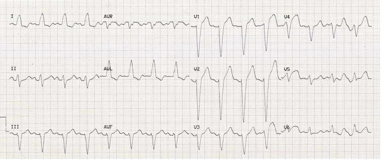
EKG not done at triage but performed at your request after evaluating patient:

image


THE TALK

Definitions

  • Hyperkalemia = potassium level > 5.5 mEq/L (a/w repolarization abnormalities)
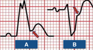
    • Peaked T waves
  • Moderate hyperkalemia = serum potassium > 6.0 mEq/L (a/w progressive paralysis of the atria)
    • P wave widening/flattening
    • PR segment prolongation
    • Eventual disappearance of P waves
  • Severe hyperkalemia = serum potassium > 7.0 mEq/L (a/w conduction abnormalities and bradycardia)
    • Prolonged/bizarre QRS morphology
    • High-grade AV block with slow junctional or ventricular escape rhythms
    • Conduction blocks
    • Sinus bradycardia/slow AF
    • Eventual development of a sine wave appearance
  • Serum potassium level of > 9.0 mEq/L can cause cardiac arrest
    • Asystole
    • Ventricular fibrillation
    • PEA with a bizarre, wide complex rhythm
  • In individual patients, serum potassium level may NOT always correlate with the EKG changes

How do I treat hyperkalemia?

  • First step: Antagonize the cardiac membrane effects of potassium (with calcium)
    • Hypocalcemia can increase the cardiotoxicity of hyperkalemia
    • Effect of calcium begins within a few minutes and lasts for ~30-60 minutes
    • Indicated only when patient has EKG changes
    • Should be administered as infusion given short half-life
  • Second step: Drive the extracellular potassium into the cells (insulin with glucose, sodium bicarbonate, and beta-2 adrenergic agonists)
    • Insulin enhances the activity of the sodium-potassium-ATPase pump in skeletal muscle
      • Effects begins w/in 10-20 minutes, peaks at 30-60 minutes and last for 4-6 hours
      • Glucose is usually given with insulin to prevent hypoglycemia
    • Beta-2-adrenergic agonists (ex: albuterol)- similar to insulin
      • Beta-2-adrenergic receptors in skeletal muscle also activate the inwardly directed Na-K-2Cl cotransporter
      • Additive effect when used with insulin and glucose.
    • Sodium bicarbonate raises serum pH, resulting in hydrogen ion release from the cells -> accompanying potassium movement into the cells
  • Third step: Remove potassium from the body
    • Loop/thiazide diuretics
      •  Limited data to support their use but makes sense clinically to give them if pt not in renal failure
    • Cation exchange resin
      • A/w intestinal necrosis even after one dose (esp in post-op patients, those with ileus, or bowel obstruction)
      • Should only be considered in patients with severe hyperkalemia and ESRD when dialysis is not available
    • Dialysis
  • In a hemodialysis patient, treating hyperkalemia acutely with these medications could drive potassium into the cells, which could then diminish subsequent potassium removal during the dialysis session if performed right afterwards, leading to rebound hyperkalemia after dialysis

TAKE HOME POINT: If a triage ECG is not done for your dialysis patient, that 10 second strip of paper should be at the top of your priority list.


REFERENCES

Williams L. “EKG of the block: Hyperkalemia” Jacobi Medical Center. Jacobi/Montefiore Emergency Medicine Conference. Bronx. November 2015. Case presentation

“ECG Features of Hyperkalaemia.” Burns, Edward. Life in the Fast Lane Medical Blog. LITFL, Web.

“Treatment and Prevention of Hyperkalemia in Adults.” Mount, David. UpToDate, Web.

aVR

Topic inspired by Dr. Jordan Smedresman’s (EM-3) ECG of the Block, November 4th 2015.

aVR

Your patient in room 11 in the green zone has chest pain/shortness of breath + ST segment elevation (STE) in lead aVR and diffuse ST segment depression (STD).

STE in aVR can be indicative of:

  1. Left main coronary artery (LMCA) occlusion/critical stenosis
  2. Proximal LAD occlusion/critical stenosis
  3. Triple vessel disease

Or:

  1. Anything that can cause severe global ischemia; GI bleed, massive PE, SVT, ect.

Treatment considerations:

  • The intervention for 2 of the 4 situations above (left main and triple vessel disease) needs coronary artery bypass grafting (CABG). If you’ve given Clopidogrel as part of your treatment, their bleeding risk during surgery has skyrocketed because of prolonged platelet inhibition.
  • STE in aVR > 1mm was a strong predictor of severe left main/3 vessel disease. These patients need a CABG and should not receive platelet inhibitors (Kosuge 2011).

Physiology:

  • The occlusion versus stenosis differentiation is an important one. Dr. Smith makes the point that, “it really just signifies widespread and diffuse subendocardial ischemia.” Left main artery OCCLUSION is usually fatal.
  • STE in aVR is not a STEMI in lead aVR; rather the STE in aVR is a reciprocal change to a leftward and inferior ST depression axis caused by diffuse subendocardial ischemia (notice the diffuse ST depression in the EKG above).

Einthovens

 Normal Variants; i.e. demand ischemia.

  • STE in aVR in presence of a supraventricular tachycardia.

SVT

  • Severe HTN can produce STE in aVR. Common in patients with bad left ventricular hypertrophy.
  • STE in aVR can also be caused by any cause of severe generalized global ischemia; severe anemia (e.g. GI Bleeding), type A dissection, massive PE, carbon monoxide poisoning etc.
  • Can also be a normal variant in left bundle branch block (LBBB)

Treat the oxygen supply and demand issue prior to diagnosing acute coronary syndrome. Reassess. If persistent ischemia, then it’s ACS. Sending a patient with acute pulmonary edema to the cath lab while they should be in the resuscitation bay on bilevel positive airway pressure and a nitroglycerine drip likely not best for your patient. If blood pressure and oxygenation controlled, and still with an ischemic ECG, then send to cath lab.

REFERENCES:

#FOAMed

Burns, Edward, ‘ST Elevation in aVR – LMCA Occlusion’ Life in the Fast Lane.

Rezaie, Salim, ‘Lead aVR: The Forgotten 12th Lead’ Academic Life in Emergency Medicine. June 20th 2013

Dr. Smith’s ECG Blog ‘STE in aVR and diffuse ST depression: It can be ACS or demand ischemia. If ACS, either posterior STEMI or subendocardial ischemia!’ November 16, 2015

Dr. Smith’s ECG Blog ‘ST Elevation in Lead aVR, with diffuse ST depression, does not represent left main occlusion’ August 2, 2014

ECG Weekly – Amal Mattu’s Case of the Week, November 9, 2015

Scholarly articles: 

Kosuge M, Ebina T, Hibi K, et al. An early and simple predictor of severe left main and/or three-vessel disease in patients with non-ST-segment elevation acute coronary syndrome. Am J Cardiol. 2011 Feb 15;107(4):495-500.

Kosuge M, Uchida K, Imoto K, et al. An early and simple predictor of severe left main and/or three-vessel disease in patients with non-ST-segment elevation acute coronary syndrome. J Am Coll Cardiol. 2015 Jun 16;65(23):2570-1.

Nikus KC, Eskola MJ. Electrocardiogram patterns in acute left main coronary artery occlusion. J Electrocardiology. 2008 Nov-Dec;41(6):626-9.

Nikus K, Pahlm O, Wagner G, et al. Electrocardiographic classification of acute coronary syndromes: a review by a committee of the International Society for Holter and Non-Invasive Electrocardiology. 2010 Mar-Apr;43(2):93, 97-98.

Rokos IC, French WJ, Mattu A, et al. Appropriate cardiac cath lab activation: optimizing the electrocardiogram interpretation and clinical decision making for acute ST-elevation myocardial infarction. Am Heart J. 2010 Dec; 160(6):995-1003.

Taglieri, N., Marzocchi, A., Saia, F., et al. Short- and Long-Term Prognostic Significance of ST-Segment Elevation in Lead aVR in Patients With Non–ST-Segment Elevation Acute Coronary Syndrome. Am J Cardiol. 2011.

Wagner GS, Macfarlane P, Wellens H, et al. AHA/ACCF/HRS recommendations for the standardization and interpretation of the electrocardiogram: Part VI: acute ischemia/infarction: a scientific statement from the American Heart Association Electrocardiography and Arrhythmias Committee, Council on Clinical Cardiology; the American College of Cardiology Foundation; and the Heart Rhythm Society. J Am Coll Cardiol. 2009 Mar 17;53(11):1007.

Williamson K, Mattu A, Plautz CU, et al. Electrocardiographic applications of lead aVR. Am J Emerg Med. 2006 Nov;24(7):864-74.


Sgarbossa’s Criteria

Topic inspired by Dr. Eliott Lee’s (EM-3) ECG of the Block, October 21st 2015.

penguinsAt the end of the day: How do I know if my patient with a left bundle branch block (LBBB) is having an acute myocardial infarction (AMI)?

  • Sgarbossa A: Concordant ST segment elevation in any lead = AMI
  • Sgarbossa B: Concordant ST segment depression in V1, V2, or V3 = AMI
  • Revised Sgarbossa C: Discordant ST segment > 25% size of R or S wave = AMI [pending validation study]

Hold on, what’s a left LBBB? A powerful leftward force; on ECG you should see several criteria:

Tintinalli:

  • Prolonged QRS duration (>0.12 s)
  • Small R wave followed by deep S wave-leads II, III, aVF, and V1-V3.
  • Large and wide R waves-leads 1, aVL, V5 and V6.
  • No Q waves in leads 1, V5, and V6.

Tintinalli, however, does not mention that there should also be:

  • Appropriate discordance: When the QRS points up, the ST segment should be a little bit depressed. When the QRS points down, the ST segment should be a little elevated. Recognizing the appropriate discordance is key to not missing an MI. Click below to enlarge.

Screen Shot 2015-10-28 at 12.52.22 PMA LBBB refresher from Life in the Fast Lane: http://lifeinthefastlane.com/ecg-library/basics/left-bundle-branch-block/


Relatively recent changes: A new LBBB does not predict high likelihood of AMI (Kontos 2011, Jain 2011, Chang 2009). Thus, in the 2013 American College Cardiology (ACC)/American Heart Association (AHA) STEMI guidelines, a new LBBB (or presumed new) is no longer an indication for cath lab activation or lytics (O’Gara 2013).

So when should my patient with a LBBB go to the cath lab? A patient with cardiopulmonary symptoms + LBBB → cath lab/lytics if:

  • Hemodynamically unstable or acute heart failure, OR
  • Concordant ST segment changes (Sgarbossa rules A or B), OR
  • ST:S ratio ≤ -0.25. In other words, discordant ST deviation ≥ 25% of the size of R or S (Smith 2012, Cai 2013). [needs validation]

  • Sgarbossa A: Concordant ST elevation ≥ 1mm in any lead = AMI
  • Sgarbossa B: Concordant ST depression ≥ 1mm in V1, V2, or V3 = AMI

Screen Shot 2015-10-28 at 12.48.28 PM

  • Revised Sgarbossa C: Discordant ST deviation ≥ 25% of the size of R or S = AMI. (Pending validation)

Screen Shot 2015-10-28 at 12.44.27 PMMore from Dr. Smith, lead author of modified Sgarbossa: http://hqmeded-ecg.blogspot.com/2012/09/modified-sgarbossa-rule-published_4.html


Screen Shot 2015-10-28 at 12.50.29 PMTo paraphrase Mattu, you can replace the 3rd step (Sgarbossa score ≥ 3) with Sgarbossa A or B. In other words, in place of using the score, if Sgarbossa A -> Primary PCI or lytics; or if Sgarbossa B -> Primary PCI or lytics. If neither, move on to looking for revised Sgarbossa C (T:S ratio ≤ -0.25, i.e. , discordant ST deviation ≥ 25% of the size of R or S (Smith 2012, Cai 2013). [needs validation]


Amal Mattu’s Sgarbossa Case:


References:

Largely adapted from Amal Mattu’s EKG weekly June 1, 2015

Cai Q, Mehta N, Sgarbossa EB, et al. The left bundle-branch block puzzle in the 2013 ST-elevation myocardial infarction guideline: From falsely declaring emergency to denying reperfusion in a high-risk population. Are the Sgarbossa Criteria ready for prime time? American Heart Journal. 2013;166(3):409–413.

Chang AM, Shofer FS, Tabas JA, et al. Lack of association between left bundle-branch block and acute myocardial infarction in symptomatic ED patients. Am J Emerg Med 2009;27(8):916–21.

Jain S, Ting HT, Bell M, et al. Utility of left bundle branch block as a diagnostic criterion for acute myocardial infarction. Am J Cardiol 2011;107(8):1111–6.

Kontos MC, Aziz HA, Chau VQ, et al. Outcomes in patients with chronicity of left bundle-branch block with possible acute myocardial infarction. American Heart Journal 2011;161(4):698–704.

Neeland IJ, Kontos MC, de Lemos JA. Evolving considerations in the management of patients with left bundle branch block and suspected myocardial infarction. J Am Coll Cardiol 2012;60(2):96–105.

O’Gara PT, Kushner FG, Ascheim DD, et al. 2013 ACCF/AHA Guideline for the Management of ST-Elevation Myocardial Infarction. JAC 2013;61(4):e78–e140.

Piktel, Joseph S. (2011). Tintinalli’s Emergency Medicine, A Comprehensive Study Guide. (7th Ed.). New York, NY: McGraw Hill.

Smith SW, Dodd KW, Henry TD, et al. Diagnosis of ST-Elevation Myocardial Infarction in the Presence of Left Bundle Branch Block With the ST-Elevation to S-Wave Ratio in a Modified Sgarbossa Rule. Ann Emerg Med. 2012;60(6):766–776.