Heart Failure…in a child?


Follow-up Rounds
5/26/2017
Article inspired by: Dr. Jeremy Price, PGY-3


THE CASE

3 month old female BIBEMS for poor feeding x 2 days as per the mother. One week ago, the mother noticed patient occasionally falls asleep while feeding. On ROS, mom has noticed occasionally increased work of breathing. Over last 12 hours, patient drank 1 ounce of formula 6 hours ago and is refusing breast feeding. Mom endorses normal wet diapers/stools and denies any fevers, vomiting or diarrhea.

Physical Exam:
Triage Vitals: HR: 158, RR: 60, SpO2: 100%, Temp: 99.5F (rectal), Weight: 6.3kg (50% percentile)

  • General: NAD, Well-appearing infant, Smiling at provider
  • HEENT: NC/AT, Anterior fontanelle open and flat, Posterior oropharynx wnl
  • Chest: CTA bilaterally, No wheezing/rales
  • Cardiovascular: RRR, No murmurs/rubs/gallops
  • Abdomen: Soft, NT/ND, No palpable masses, No organomegaly
  • GU: Normal female genitalia
  • Skin: Intact, no rashes
  • Neuro: Alert, Awake, Symmetric face, Normal tone


Critical Intervention:
-Baby appeared well and was observed during a feed before deciding disposition.

-During the feed, patient appeared diaphoretic and only took 1 ounce before refusing any more formula.

-After the feed, patient noted to be tachypneic in 60s-70s, with retractions
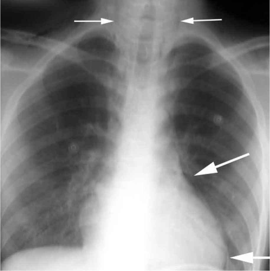
CXR Interpretation:
Markedly enlarged cardiac silhouette. The lungs are enlarged and the vascularity is increased

Follow-Up Studies:


  • TTE: showing severely dilated LV with EF 13%, Rest of heart structurally normal, with normal coronary origins, no coarctation of aorta
  • Pro-BNP: 73,926
  • Troponin: 0.35

THE TALK

The presentation of pediatric heart failure varies drastically based on its cause and is different from the classic presentation of heart failure we are used to in the adult population.

What are some signs I should look for on my physical exam to suspect heart failure?

  • Tachycardia
  • Decreased perfusion (cool/mottled extremities, decreased capillary refill, low BP)
  • S3 gallop
  • Respiratory distress (tachypnea, retractions, grunting)
  • Systemic congestion (hepatomegaly, splenomegaly, ascites, peripheral edema)
  • High BP in upper extremities (suggestive of coarctation of aorta)
  • Weak pulses in lower extremities (suggestive of coarctation of aorta)
  • Palpable “thrill” (suggestive of a shunt lesion)
  • “Heave” or laterally displaced point of maximal pulse (suggestive of long-standing cardiomyopathy)

Is there any system I can use to classify heart failure similar to the NYHA classification in adults?

Modified Ross Heart Failure Classification

  • Class I: No limitations or symptoms
  • Class II:
    • Infants: Mild tachypnea or diaphoresis with feeding
    • Older children: Mild to moderate dyspnea on exertion
  • Class III:
    • Infants: Growth failure and marked tachypnea or diaphoresis with feeding
    • Order children: Marked dyspnea on exertion
  • Class IV: Symptoms as rest (tachypnea, retractions, grunting or diaphoresis)

Okay, what tests can I order to confirm my diagnosis?

  • CXR (to assess for cardiomegaly and pulmonary congestion)
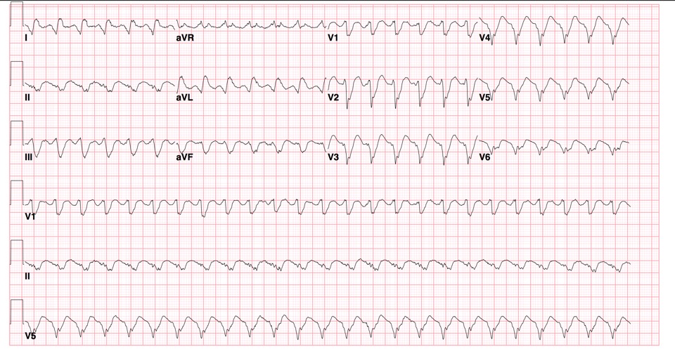
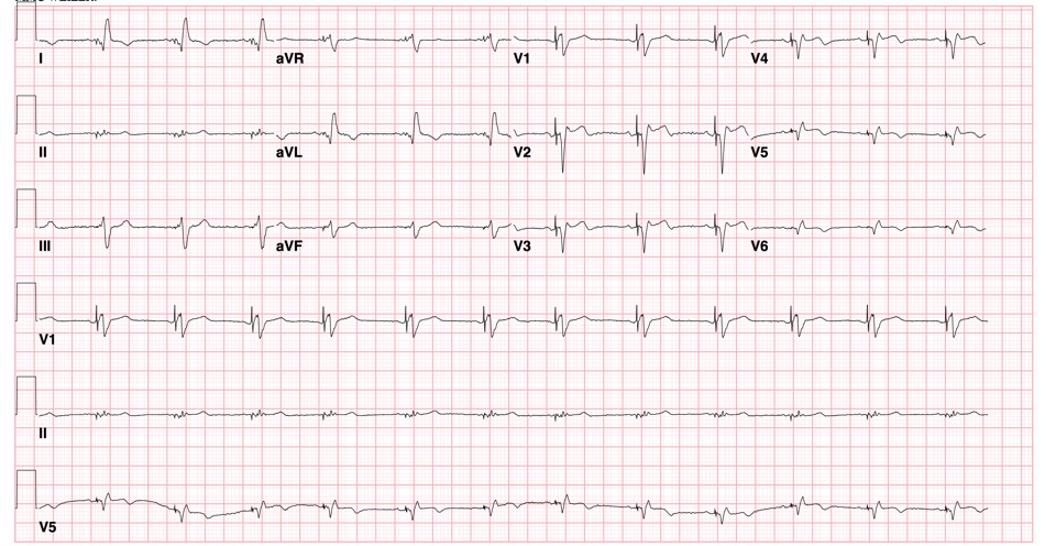
  • EKG (ST segment and T wave abnormalities are common in cardiomyopathy)
  • Echocardiogram (to assess anatomy, ventricular size and function)
  • Lab Tests
    • Chem, including LFTs
    • CBC (to r/o anemia as an exacerbating factor)
    • Pro-BNP
    • Troponin

This was too long and I didn’t read it- what should I know?

  • If you have a pediatric patient with a feeding complaint who looks amazing in your ED, watch the patient during a feed to see what the patients are talking about!

REFERENCES/FURTHER READING:

  • Singh, Rakesh K., and TP Singh. “Heart Failure in Children: Etiology, Clinical Manifestations, and Diagnosis.” UpToDate. 21 Apr. 2017. Web.
  • Singh, Rakesh K., and TP Singh. “Heart failure in children: Management.” UpToDate. 21 Apr. 2017. Web.
  • Jayaprasad, N. “Heart Failure in Children.” Heart Views : The Official Journal of the Gulf Heart Association. Medknow Publications & Media Pvt Ltd, Sept. 2016. Web.

TRALI

Calvin D. Sun, MD, PGY-3
Follow-Up Rounds 5/12/2017


THE CASE

Triage Vitals: T 97.4F,  BP 137/49, HR 66, RR 18, O2 100% on RA

CC: Lower Back Pain, B/L LE numbness

HPI:

  • 56M
  • No known PMH
  • Lower back pain radiating to BL feet x 1 week
  • Numbness of BL feet since x 1 day
  • Denies fever, LOC, headache, trauma, history of abdominal surgeries, abdominal pain
  • Jehovah’s Witness
  • Nightly AM Advil at home for back pain/sleeping aid also relieves pain.
  • Pt also c/o blood on top of brown stool x months
  • Normal colonoscopy and EGD 3 years ago

PHYSICAL EXAM

  • Gen: AAO x 3, well appearing, NAD
  • HEENT: atraumatic, no cyanosis, no pallor, MMM
  • CV: RRR, no murmurs appreciated
  • Pulm: CTA B/L, no wheezing
  • Abd: S, NT, ND, + BS in all 4 quadrants
  • Extr: pulses palpable bilaterally, no edema
  • Neuro: moving all 4 extremities, no facial droop, no focal deficits, CN II-XII grossly intact

STUDIES

CBC
WBC (/nL) 10.2 (3.5 – 11.0)
Hgb (g/dL) 4.4 (12.0 – 16.0)


Coags
aPTT (sec) 23.3 (20.1 – 31.2) sec
PT 13.4 (9.5 – 12.2) sec
INR 1.0 (0.9 – 1.2)


CMP
Na 139 (135 – 145) mEq/L
K 7.1 (3.5 – 5.0) mEq/L
Cl 112 (98 – 108) mEq/L
CO2 17 (24 – 30) mEq/L
BUN 18 (5 – 26) mg/dL
Creat 2.27 (0.1 – 1.5) mg/dL
Gluc 111 (70 – 105) mg/dL

INTERVENTIONS

  • Patient begins 1u pRBC transfusion
  • 5 minutes later and after receiving 100 mL of blood, patient begins to feel SOB

INTERVENTIONS – 5 MINUTES LATER

  • Nurse notices acute respiratory distress
  • Patient becomes altered, obtunded, not answering questions, diaphoretic w/ retractions, unable to speak
  • PO2 on NRB mask 79%
  • Patient intubated
  • …and pRBC transfusion stopped

Portable CXR


TRALI

  • Transfusion-related acute lung injury
  • Incidence among patients receiving transfusion
    • 04-0.10% (1 in 5000) in all cases
    • 5-8% in critically ill patients
  • During or within 6 hours after blood product administration
  • Can occur with any blood product
  • Being critically ill is the highest risk factor
  • Pathogenesis (2-hit):
    • Neutrophils primed to respond
    • Neutrophil activation by a factor in the blood product → damage capillary endothileum in lung → pulmonary edema

CLINICAL PRESENTATION OF TRALI

  • Hypoxemia (100%)
  • Bilateral pulmonary infiltrates (100%)
  • Pink frothy airway secretions (56%)
  • Fever (33%)

DIFFERENTIALS RELATED TO TRALI

  • TACO: Transfusion-associated circulatory overload
    • More associated with volume overload
  • Hemolytic transfusion reaction
    • More associated with fever and chills
  • Anaphylaxis
    • More associated with stridor, cough, wheezing
  • Sepsis
    • More associated with fever and hypotension with evidence of an active infectious process

MANAGEMENT OF TRALI

  • O2, Airway management, CXR
  • Stop transfusion immediately
    • Return blood product to the blood bank for a transfusion reaction workshop (CBC, bilirubin, haptoglobin, Coombs)
  • Supportive care
    • Intubation often required (70-80%)
  • Mixed results with steroids; not recommended
  • Patients appear not to be at increased risk for repeat episodes with future transfusions if from other donors

REFERENCES

Fung YL, Silliman CC. The role of neutrophils in the pathogenesis of transfusion-related acute lung injury. Transfus Med Rev 2009; 23:266.

Silliman CC. The two-event model of transfusion-related acute lung injury. Crit Care Med 2006; 34:S124.

Bux J, Sachs UJ. The pathogenesis of transfusion-related acute lung injury (TRALI). Br J Haematol 2007; 136:788.

van Stein D, Beckers EA, Sintnicolaas K, et al. Transfusion-related acute lung injury reports in the Netherlands: an observational study. Transfusion 2010; 50:213.


VT Storm

Brian Gilberti, MD, PGY-3
Follow-Up Rounds 3/3/2017

CASE
Triage Vitals: T 98.0, HR 152, BP 66/48, RR 20, SpO2 97%
CC: Dizziness

HPI
  • 75 yo M
  • PMH CAD s/p CABG, HFrEF (25%, AICD), HTN, DM
  • Worsening over past two days
  • Denied fevers, vertigo, CP, SOB
  • Compliant with medications
PHYSICAL EXAM
Vitals: HR 152, BP 66/48
General: In no acute distress
HEENT: no JVD
CV: Tachycardic, no murmurs
Pulm: Mild rales at bases bilaterally
Abd: Soft, NT
Extremities: Trace pitting edema

STUDIES

ECG on presentation

ECG 3 months prior

Labs: Unremarkable
ICD Interrogation

  • Episodes
    • Today: VT
    • 2 days prior: VT >18 hrs
    • 3 days prior: VT > 18 hrs

Definition

  • Without ICD
    • ≥2 episodes of VT within 24 horus
    • Recurrence of VT within 5 mins of termination of prior episode
    • >24 hour episode
  • With ICD
    • ≥3 episodes requiring device intervention
  • Incidence 2-10% per year in patients with ICDs


Triggers

  • Structural abnormality
  • Myocardial ischemia
  • Heart failure
  • Thyrotoxicosis
  • Hypomagnesemia
  • Hypokalemia
  • QT prolongation


Indicators of Ventricular Tachycardia on ECG (vs SVT with aberrancy)

  • Regular
  • Northwest axis (-90 to +/-180)
  • Compared to sinus rhythm, axis shift >40 during wide-complex tachycardia
  • QRS duration >160 msec
  • Negative concordance of QRS morphology in V1-V6
    • Positive concordance as well, but not as strongly suggestive of VT
  • Fusion beats (diagnostic of VT)


Treatment

  • Unstable
    • Cardiovert
  • Stable
    • Antiarrhythmics
      • Procainamide
        • Procainamide was more effective than Amiodarone in terminating VT within 20 mins (67% vs 38%) and had fewer MACE (9% vs 41%).1
          • NB: This was not a study of pts with electrical storm
        • AHA Class IIa for stable monomorphic VT
        • Contraindicated in patients with impaired renal function because its active metabolite, N-acetylprocainamide, is excreted by the kidneys
        • Administer until:
          • Max dose of 17 mg/kg or 1 g
          • Arrhythmia resolves
          • QRS widens by >50%
          • Pt bradycardic
          • Pt hypotensive
      • Amiodarone
        • Reduces frequency of recurrent episodes of ventricular arrhythmia2,3
        • Mexiletine/Purkinje catheter ablation4
          • Effective in relatively narrow QRS refractory to Amiodarone
        • Acute side effects5
          • Hypotension (diluent)
          • Bradycardia
          • AV block
          • QT prolongation
          • Interstitial lung disease/pulmonary fibrosis
          • Hepatitis
        • Lidocaine6,7
          • Most efficacious in ischemic myocardium
          • Conversion rates from VT = 8-30%

Beta Blockers6,7

  • Provides competitive sympathetic blockade
  • Metoprolol preferred
  • Propranolol reasonable but no studies supporting the use of one over the other
    • Use with caution in patients with HFrEF

Catheterization8

  • Electrical storm often seen in ischemic heart disease
  • Urgent cath if MI considered etiology

Catheter Ablation6,9

  • Recommended if VT persists despite amiodarone/beta blocker
  • Superior to escalating medical therapy10

References

1)    Ortiz M, Martín A, Arribas F, et al. Randomized comparison of intravenous procainamide vs. intravenous amiodarone for the acute treatment of tolerated wide QRS tachycardia: the PROCAMIO study. Eur Heart J. 2016 Jun 28.

2)    Levine JH, Massumi A, Scheinman MM, et al. Intravenous amiodarone for recurrent sustained hypotensive ventricular tachyarrhythmias. Intravenous Amiodarone Multicenter Trial Group. J Am Coll Cardiol 1996; 27:67.

3)    Scheinman MM, Levine JH, Cannom DS, et al. Dose-ranging study of intravenous amiodarone in patients with life-threatening ventricular tachyarrhythmias. The Intravenous Amiodarone Multicenter Investigators Group. Circulation 1995; 92:3264.

4)    Murata H, Miyauchi Y, Hayashi M, et al. Clinical and Electrocardiographic Characteristics of Electrical Storms Due to Monomorphic Ventricular Tachycardia Refractory to Intravenous Amiodarone. Circ J 2015; 79:2130.

5)    Long B, Koyfman A. Best Clinical Practice: Emergency Medicine Management of Stable Monomorphic Ventricular Tachycardia. J Emerg Med. 2016 Oct 14. pii: S0736-4679(16)30721-1.

6)    Zipes DP, Camm AJ, Borggrefe M, et al. ACC/AHA/ESC 2006 Guidelines for Management of Patients With Ventricular Arrhythmias and the Prevention of Sudden Cardiac Death: a report of the American College of Cardiology/American Heart Association Task Force and the European Society of Cardiology Committee for Practice Guidelines (writing committee to develop Guidelines for Management of Patients With Ventricular Arrhythmias and the Prevention of Sudden Cardiac Death): developed in collaboration with the European Heart Rhythm Association and the Heart Rhythm Society. Circulation 2006; 114:e385.

7)    Eifling M, Razavi M, Massumi A. The evaluation and management of electrical storm. Tex Heart Inst J 2011; 38:111.

8)    Authors/Task Force members, Windecker S, Kolh P, et al. 2014 ESC/EACTS Guidelines on myocardial revascularization: The Task Force on Myocardial Revascularization of the European Society of Cardiology (ESC) and the European Association for Cardio-Thoracic Surgery (EACTS)Developed with the special contribution of the European Association of Percutaneous Cardiovascular Interventions (EAPCI). Eur Heart J 2014; 35:2541.

9)    Nayyar S, Ganesan AN, Brooks AG, et al. Venturing into ventricular arrhythmia storm: a systematic review and meta-analysis. Eur Heart J 2013; 34:560.

10)   Sapp JL, Wells GA, Parkash R, et al. Ventricular Tachycardia Ablation versus Escalation of Antiarrhythmic Drugs. Circulation. N Engl J Med. 2016 Jul 14;375(2):111-21.

Epiglottitis

Follow-up Rounds
1/13/2017
Article inspired by: Dr. Maninder Singh, PGY-3


THE CASE

37 y/o M with no significant PMHx who p/w “allergic reaction” after taking the first dose of PCN 2 hours ago prescribed for Strep Throat diagnosed at an urgent care center. He endorses a sore throat and fever x 2 days but denies any hives or lip/tongue swelling. Denies any prior allergic reactions.

Physical Exam:

  • Vitals: BP: 133/76, HR: 107, RR: 15, Temp: 99F, SpO2: 100%
  • General: Appears well
  • HEENT: NC/AT, PERRL, EOMI, Uvula is midline, Moist mucous membranes, No tonsillar abscesses, Muffled voice, No visible upper oral airway obstruction, Increased saliva production noted; Neck supple, Some lymph node swelling to left precervical nodes
  • Cardiovascular: RRR, No murmurs/rubs/gallops
  • Chest: CTA b/l, No wheezes/rales/rhonchi
  • Abdominal: Soft, NT/ND, BS+ x4
  • Extremities: FROM


Soft Tissue Neck X-ray:

epiglottitis


THE TALK

What is an epiglottis?

  • Back wall of the vallecular space below base of tongue
  • Infectious epiglottitis = cellulitis of epiglottis
    • Progresses to involve entire supraglottic larynx (including aryepiglottic folds and arytenoids) leading to difficulty breathing

Isn’t epiglottitis a pediatric diagnosis?

  • Incidence decreased in children since haemophilus influenza vaccination
  • Incidence in adults (2006): 1.6 cases per 100,000 adults
    • Usually a/w HTN, DM, Substance abuse or immune deficiency

How do I diagnose it?

  • Fiberoptic nasal laryngoscopy = gold standard
  • Lateral neck x-ray: look for the “thumbprint” sign
  • CT neck (if diagnosis unclear)

What do I do if I suspect/diagnose it?

  • MANAGE AIRWAY- Be prepared for a cricothyrotomy!
  • Have patient sitting up in bed
  • Consider prophylactic intubation
    • Risk of laryngospasm with scope
  • Heliox (mixture of Helium and Oxygen) can be used as a temporizing measure
  • Augmentin or Ampicillin-sulbactam (Unasyn) are preferred initial antibiotics
  • Consider Vancomycin if patient is critically ill and MRSA a possible etiology
  • NSAIDS for pain control
  • +/- Steroids for symptomatic relief

This was too long and I didn’t read it- what should I know?

  • Patient with sore throat and muffled voice but no obvious signs of upper airway obstruction need further work up
  • Have your triple set up ready (direct laryngoscope, video laryngoscope and cricothyroidectomy) and have backup (ENT/Anesthesia) available for a difficult airway

REFERENCES/FURTHER READING:

  • Frantz TD, Rasgon BM, Quesenberry CP. Acute Epiglottitis in Adults: Analysis of 129 Cases. 1994;272(17):1358-1360.
  • Woods, Charles. “Epiglottitis (supraglottitis): Clinical Features and Diagnosis.” UpToDate, 23 June 2015. Web.
  • Rogers, Matt. “Epiglottitis.” Core EM, 26 Aug. 2015. Web.

Boerhaave Syndrome

Case presented by Dr. Jordan Smedresman
Follow-up Rounds 11/6/2015

CASE
Triage Vitals: T 98.0, HR 92, BP 150/102, RR 20, SpO2 92%
CC: Back pain

HPI
  • 72 yo M with PMH HTN BIBEMS for L sided back pain
  • Started after eating
  • Denied antecedent vomiting
  • Pain associated with dyspnea
PHYSICAL EXAM
Vitals: SpO2 88% on RA, 96% on 2L NC, BP 146/94
General: In acute distress
CV: RRR, no murmurs
Pulm: Diminished breath sounds on L

Abd: Soft, NT

STUDIES
EKG: NSR, no TWIs or ST deviations
POCUS: Possible L-sided pneumothorax
Labs: Unremarkable
CXR: L-sided hydropneumothorax, pneumomediastinum
CT Thorax: Boerhaave syndrome with L-sided pneumothorax, extensive pneumomediastinum


BACKGROUND
  • From sudden increase in esophageal pressure/decrease in intrathoracic pressure
    • Vomiting
    • Childbirth
    • Seizure
    • Prolonged coughing/laughing
    • Weightlifting
  • 15% of esophageal perforations
    • Most iatrogenic > FB or malignancy
  • Most common location of perforation: L posterolateral aspect of distal intrathoracic esophagus
  • Gastric contents in mediastinal cavity → chemical mediastinitis → bacterial infection
  • Pleural cavity may be violated from inflammation or initial perforation
  • ~100% mortality if untreated
CLINICAL MANIFESTATIONS
  • Symptoms (% of pts)
    • Chest pain (89%)
    • Dyspnea (67%)
    • Dysphagia (3%)
    • Neck pain (11%)
    • Neck swelling (6%)
    • Hoarse voice (6%)
  • History of retching (NB: 25-45% don’t have history of vomiting)
  • Crepitus with palpation of chest wall
  • Hamman’s sign: mediastinal crackling with heartbeat
  • Within hours:
    • Odynophagia, dyspnea, mediastinitis, sepsis
DIAGNOSIS
  • CXR
    • Not sensitive; may require hours for signs to develop
    • Findings
      • Mediastinal/free peritoneal air/SQ emphysema
      • Pleural effusion
      • Mediastinal widening

screen-shot-2016-11-29-at-4-51-24-pm

  • CT
    • Findings
      • Esophageal wall edema/thickening
      • Mediastinal widening
      • Air/fluid in pleural spaces/retroperitoneum

screen-shot-2016-11-29-at-4-50-53-pm

MANAGEMENT
  • NPO
  • Broad spectrum abx
  • Protonix gtt
  • CT Surgery consult
    • Surgical candidates:
      • Diffuse extravasation
      • Extension of perforation
      • Sepsis
      • Progression of pneumomediastinum or pneumothorax
      • Patients with empyema

REFERENCES

Blencowe NS, Strong S, Hollowood AD. Spontaneous oesophageal rupture. BMJ 2013;346:f3095.

Carrott PW, Jr., Low DE. Advances in the management of esophageal perforation. Thorac Surg Clin 2011;21:541-55.

Henderson JA, Peloquin AJ. Boerhaave revisited: spontaneous esophageal perforation as a diagnostic masquerader. Am J Med 1989;86:559-67.

Newcomb AE, Clarke CP. Spontaneous pneumomediastinum: a benign curiosity or a significant problem? Chest 2005;128:3298-302.

Triadafilopoulos G. “Boerhaave syndrome: Effort rupture of the esophagus.” Up To Date. http://www.uptodate.com, 26 Apr. 2016. Web. 28 Nov. 2016. https://www.uptodate.com/contents/boerhaave-syndrome-effort-rupture-of-the-esophagus

Pheochromocytoma

Inspired by Dr. Serene Rich
Follow-Up Rounds 2/19/2016

Case
  • 27 yo F BIBEMS for AMS
  • Progressively altered and emotionally labile over past week, per family
  • No PMH/PSH/Medications
Physical Exam
Vitals BP 163/129, HR 123, RR 18, O2 98%
General: AAOx1 (person), NAD, emotionally labile
HEENT: NC/AT, no thyroid nodules, PERRL, EOMI
CVS: Tachycardia, regular rhythm, S1, S2
Pulm: CTAB

Abd: Soft, NT, ND

Studies
CBC/BMP, U/A unremarkable
bHCG neg
TSH 0.14, T4 0.85
Trop 0.180
Utox neg
EKG: Sinus tachycardia, TWIs in lateral leads, no ST deviations

NCHCT: No acute findings
CT A/P: Adrenal mass c/w pheochromocytoma

Course: Patient with likely hypertensive encephalopathy/demand ischemia due to catecholamine excess. Admitted to CCU. 24-urine with elevated metanephrines. Treated with cyclobenzamine and nicardipine; started on propranolol on HD 2.


PHEOCHROMOCYTOMA

Background
  • Catecholamine-secreting tumor
  • Occur in 0.2% of patients with hypertension
  • Age: 30-40s
  • Associated syndromes: VHL, MEN2, NF1
  • “Rule of 10’s”
    • 10% bilateral
    • 10% extra-adrenal
    • 10% familial
    • 10% malignant
  • Symptoms caused by hypersecretion of one or combinations of norepinephrine/epinephrine/dopamine
Presentation
  • Classic triad: Episodic HA, sweating, tachycardia
    • Most do not have all three
    • HA occurs in 90% of symptomatic patients
    • Sweating occurs in 60-70%

Screen Shot 2016-03-17 at 11.30.35 AM

  • Paroxysmal HTN most common sign
  • Some patients may present hypotension that may be 2/2:
    • Volume depletion
    • Abrupt cessation of excessive catecholamine secretion
    • Desensitization of adrenergic receptors
    • Hypocalcemia
  • Paroxysms may be triggered by:
    • Micturition (urinary bladder pheochromocytoma)
    • Drugs: Glucagon, IV contrast, tyramine, reglan
Diagnosis
  • Biochemical testing
    • Should be performed in all patients with suspected pheochromocytoma

Screen Shot 2016-03-17 at 11.31.18 AM

  • Imaging
    • CT A/P
      • Detects almost all sporadic tumors
      • Sn = 98-100%, Sp = 70%
      • Consider using low-osmolar contrast as contrast may induce a paroxysm 
      • In ED setting, this will typically precede biochemical testing, though biochemical testing is recommended as first step in work-up of suspected pheo
        • NB: 95% are within abdomen, 10-15% extra-adrenal tumors
    • Screen Shot 2016-03-17 at 3.16.42 PMMIBG
      • May detect tumors not seen on CT/MRI
      • Performed in patients with pheochromocytomas >10 cm due to increased risk of malignancy
Treatment
  • Alpha blockade
    • Phenoxybenzamine preferred agent
  • Beta blockade
    • Should be started only after alpha blockade initiated due to concern for unopposed alpha
    • Propranolol may be used
  • Calcium Channel Blockers
    • Typically used as additional agent after alpha blockade
    • May be used as single agent
    • Nicardipine typically used
  • Surgery
    • Definitive treatment is lap adrenalectomy

Lenders JW, Eisenhofer G, Mannelli M, Pacak K. Phaeochromocytoma. Lancet 2005;366:665-75.

Young WF, Jr. Clinical practice. The incidentally discovered adrenal mass. N Engl J Med 2007;356:601-10.

Young, Williams. “Clinical presentation and diagnosis of pheochromocytoma.” Up To Date. http://www.uptodate.com, 24 Feb. 2015. Web. 16 Mar. 2016. http://www.uptodate.com/contents/clinical-presentation-and-diagnosis-of-pheochromocytoma

Young, Williams. “Treatment of pheochromocytoma in adults.” Up To Date. http://www.uptodate.com, 27 Oct. 2015. Web. 16 Mar. 2016. http://www.uptodate.com/contents/treatment-of-pheochromocytoma-in-adults


Aortic Dissection

Follow-up Rounds
1/22/2016
Article inspired by: Dr. Joshua Schwarzbaum, PGY-3


THE CASE

60 y/o F PMHx HTN BIBEMS after bystanders called for erratic behavior. As per triage nurse, patient is rambling while speaking and diaphoretic but with no visible injuries. Patient reports he cannot breathe and that he was told he passed out while walking his dog. He reports he feels cold and sweaty. Denies fevers, chills, nausea, vomiting, chest pain, abdominal pain, or recent travel.

EKG- sinus tachycardia

Physical Exam:
Vitals: BP- 106/74, HR-107, RR-20, Temp-96F, SpO2- 94%, FS- 125
General: Mild distress, lying in bed
Chest: CTA b/l
Cardiovascular- S1/S2, Tachycardic
Abdomen- Soft, NT/ND
Extremities- Warm, No peripheral edema
Neurological- Waxing/waning mental status, PERRL, Moving all extremities, Will not comply with exam

Labs:
138/hemolyzed/108/17.4/14/1.5<265
15.2>15.4/46.3<184
Trop: 0.22, CK: 235

CXR: No acute cardiopulmonary process

CT Head: No acute intracranial pathology

CT Chest: Findings are consistent with bibasilar pulmonary emboli, extremely slow flow through the heart and evidence of heart failure with smooth thickening of the interstitium and geometric polygons formation. Bibasilar atelectasis/infiltrate. Moderate pericardial effusion which is hemorrhagic in nature and with a history of hypertension, rule out dissection of the aorta which is not opacified due to extremely slow flow.

Echocardiogram:


THE TALK

Is there a way to classify aortic dissections?

  • Arbitrary way: Acute (<2 weeks of symptoms) vs Chronic
    • Life threatening complications 2/2 branch involvement or aortic rupture more common in acute
  • Stanford Classification
    • Type A
      • Involving ascending aorta, regardless of site of primary intimal tear
    • Type B
      • Everything else
  • Debakey Classification
    • Type 1
      • Originating in ascending aorta and propagating to at least aortic arch
    • Type 2
      • Originating in and confined to ascending aorta
    • Type 3
      • Originating in the descending aorta and extending distally or proximally

What type of dissection is more common?

  • Ascending aortic dissection ~2x more common than descending aortic dissection
    • Right lateral wall of ascending aorta = most common site
    • Aortic arch involvement occurs in ~30%

How does an aortic dissection happen?

  • Primary event = tear in aortic intima
  • Blood passes into aortic media through tear, creating false lumen
  • Related to shear forces

How can people die from aortic dissection?

  • Rupture of dissection into pericardium leading to cardiac tamponade
  • Acute dissection of aortic valvular annulus leading to severe aortic regurgitation
  • Obstruction of coronary artery ostia leading to MI
  • Abdominal aortic branch vessel obstruction leading to end organ failure

What are some risk factors for aortic dissection?

  • HTN (abrupt, transient, severe increase in BP, ex: crack cocaine or high intensity weight lifting)
  • Collagen disorders (ex: Marfan, Ehler-Danlos, annuloaortic ectasia)
  • Preexisting aortic aneurysm
  • Bicuspid aortic valve (always involves ascending aorta)
  • Aortic instrumentation/cardiac surgery
  • Aortic coarctation
  • Turner syndrome
  • Vasculitis (ex: Giant cell arteritis, Takayasu arteritis, RA, Syphilitic aortitis)
  • Trauma (but usually causes aortic rupture or transection)
  • Pregnancy/Delivery

What are some things I should elicit on my H&P?

  • Acute pain (typically severe, sharp/knife-like)
    • Abdominal pain/Mesenteric ischemia (think celiac or mesenteric arteries involvement)
    • Back or flank pain/Renal failure (think renal artery involvement)
  • Pulse deficit (a weak or absent carotid, brachial, or femoral pulse resulting from the intimal flap or compression by hematoma)
    • Upper extremity pulselessness/Hypotension (think subclavian artery involvement)
    • Lower extremity pain/pulselessness/weakness (think common iliac artery involvement)
  • Heart murmur (new diastolic decrescendo murmur)
    • Aortic insufficiency/Heart failure (think aortic valve involvement)
  • Focal neurologic deficit
    • Stroke/Syncope (think brachiocephalic, common carotid or left subclavian artery involvement)
    • Paraplegia (think intercostal artery involvement as they give off spinal/vertebral artery)
    • Horner syndrome (ptsosis/miosis/anhidrosis) (think superior cervical sympathetic ganglion involvement)
  • Hypotension
    • Cardiac tamponade (think pericardium involvement)
    • MI (think right coronary artery involvement)
    • Hemothorax/Hemoperitoneum (think thoracic or abdominal aorta involvement)

How do I manage someone with aortic dissection?

  • Pain control with narcotics (morphine)
  • If hemodynamically unstable/airway compromise, intubate
  • Heart rate control to <60bpm (IV beta blocker ex: propranolol or labetalol)
    • Labetalol is an alpha- and beta-receptor antagonist (may be more effective in controlling both heart rate and blood pressure as a single agent)
    • Esmolol has short half-life and can be titrated to effect
    • Esmolol also beneficial in asthma or heart failure (pts who are intolerant to BB)
    • Other options if cannot use BB: Verapamil or Diltiazem
  • Reduction of systolic blood pressure of 100-120mmHg
    • If after BB, still elevated- can use Nitroprusside (but only after BB because vasodilation alone induces reflex activation of sympathetic nervous system -> enhanced ventricular contraction -> increased aortic wall shear stress)
    • Can use ACE-inhibitors
    • AVOID HYDRALAZINE (increases aortic wall shear stress)

What if my patient is hypotense?

  • Prior to giving volume, determine cause:
    • Blood loss
    • Hemopericardium with tamponade
    • Valvular dysfunction
    • Left ventricular systolic dysfunction
  • AVOID inotropic agents (increase aortic wall shear stress)

REFERENCES

Schwarzbaum J. “Follow up Rounds: Aortic Dissection” Jacobi Medical Center. Jacobi/Montefiore Emergency Medicine Conference. Bronx. Jan 2016. Case Presentation

Manning, Warren J., and James H. Black. “Clinical Features and Diagnosis of Acute AorticDissection.” Clinical Features and Diagnosis of Acute Aortic Dissection. UpToDate, 19 Feb. 2016. Web.

Manning, Warren J. “Management of Aortic Dissection.” Management of Aortic Dissection. UpToDate, 20 Nov. 2013. Web.

Hyponatremia

Inspired by:
Joshua Scharwbaum
Follow-Up Rounds 1/22/16

THE CASE
CC: Seizure
HPI:
  • 35 yo F with no PMH BIBEMS for generalized seizure on day of presentation
  • No history of seizures
  • Lasted for 1 minute and resolved prior to EMS arrival
  • No fevers, N/V/D, SOB, CP per collateral
PMH/PSH: Denies

Medications: None

Physical Exam
Vitals: BP 140/62, HR 112, RR 12, T 98.6, O2 99%, FS 120
General: Confused, responding to verbal stimuli, NAD
CVS: Tachycardic, regular rhythm
Pulm: CTAB
Abd: Soft, NT, ND

Neuro: PERRL, opening eyes spontaneously but not interactive, nonverbal, moving all extremities

Studies:
CBC: 9.6 > 14.1/40.1 < 210
BMP: Na 115, K 4.0, Cl 69, HCO3 23, BUN 6, Cr 0.44 Glucose 105
Urine Electrolytes: Na 29 (nl: 15-267), Cl 20 (nl: 20-295), Glu 4 (nl: 0-33), K 15 (nl: 17-164), Osm 131 (nl: 300-1300)

HYPONATREMIA
Stratification:
  • Acute (<24 hrs)
  • Chronic (>24 hrs)
  • Mild: Na 130-135
  • Moderate: Na 121-129
  • Severe: <120
Symptoms:
  • Typically absent in chronic hyponatremia if Na >120
  • Mild-Mod
    • HA, N/V, fatigue, gait, confusion
  • Severe
    • Seizures, obtundation, coma, respiratory arrest
Classification:
  • Hypovolemic
    • d/t GI or renal lossess
  • Normovolemia
    • SIADH, polydipsia
  • Hypervolemia
    • Heart failure, cirrhosis
What should be considered in a patient with hyponatremia and elevated urine Na (>20 mEq/L)?
  • NB: Normal response would be to produce urine with Na <10 mEq/L
  • SIADH
  • Hypothyroidism
  • Adrenal insufficiency
  • Osmotic diuresis
  • Diuretics
  • Renal failure
How should these patients be managed in the ED?
  • Treat those with Na <115 mEq/L or when patient is symptomatic
  • Urine lytes only useful before starting treatment
  • Use hypertonic 3% saline in:
    • Na <120 mEq/L
    • Rapidly developed (>0.5 mEq/L decrease per hours)
    • a/w coma or seizure
  • Goal increase: 6 mEq/day
    • Can be increased to 6 mEq in 6h if having CNS event (Rule of 6’s)
What do I do if the patient is seizing?
  • Give patient 100 cc bolus of 3% saline over 10-60 minutes
    • Should raise Na by 2 mEq/L
  • You may give a second 100 cc bolus if patient continues to seize
What do I do if I overcorrect? (i.e. Na 14 mEq increase in 2 hours)
  • DDAVP 1-2 mg (little evidence in humans)
    • Avoid in psychogenic polydipsia
  • D5W 6 cc/kg over an hour with meds (consult renal)
“How can you blow this case?”
It is easy to blow any hyponatremia case by raising the sodium too rapidly and causing osmotic demyelination.  Giving 1-2 L of NS in a pt with an unexplained change in MS before we get back the labs is a common scenario in the ED.  Even if you have the labs back before giving NS,  it is easy to be too aggressive in correcting the sodium.  Remember that you just want to raise the sodium a small amount to the level at which they were before the change in mental status.  You do not need to raise the sodium to normal.

COMMENTS
Dr. Gruber:
“Hypertonic saline is indicated when pt is having a seizure, coma or suspected cerebral herniation. It is not indicated in pt with very low Na (i.e. <120) if there is no neurologic emergency. Sodium bicarb can be used if hypertonic saline is not available.

Assuming there is no neuro emergency, it is important to figure out the pt’s fluid status. Although it is difficult to estimate the pt volume status clinically at the bedside with any accuracy, we can look at the pt history (i.e. vomiting), HR, BP, JVD, presence of pedal edema, orthopedic VS and the point of care U/S of the IVC. This can be used to make rough determination of fluid status which dictates whether pt gets fluids, has fluid restriction and/or gets diuretics.

In the hypovolemic, hyponatremic pt, it is important to restore volume but be mindful of the sodium concentration of the fluid you use. Using Ringers lactate has a Na of 128 will likely result in slower rise serum sodium than using normal saline.

In the pt who is hypokalemic and hyponatremic, correcting the hypokalemia will help raise the sodium but may result in overcorrection of the sodium. It is important to correct hypokalemia as it is a risk factor for developing osmotic demyelination (AKA
central pontine myolysis) but it is important to factor in the contribution of supplemental K in raising Na. Other risk factors for developing OD are being elderly, malnourished state and chronic severe hyponatremia.

We talked about the Rule of 6’s in correcting Na (ie raise Na 6 points) in 6hrs if seizure or coma but raise Na 6 points in 24hrs if no neuro emergency. There is also the Rule of 100s to prevent rapid overcorrection. This rule is based on the fact that the rapid rise in Na is due to more to the pt’s free water diuresis after you give fluids than to the fluids that you are giving. Monitoring the urine output is key to preventing overcorrection of the Na. Therefore, in the pt with critically low Na, urine output and IVF need to be closely monitored:

-likely need foley
-if urine output >100cc/hr, send STAT urine osmolariy and Na
-if urine osmolarity<100, consider DDAVP
-continue to follow latter 2 steps

In terms of causes of hyponatremia, meds such as SSRIs cause SIADH and hyponatremia and chronic steroids cause adrenal insufficiency. Exercise associated hyponatremia is seen amongst endurance athletes (i.e. marathon runners). Their hyponatremia results from taking in more free water than they are able to clear in their urine. One can blow the case by assuming that their change in MS or other complaints are due to dehydration rather than hyponatremia.

Pts taking ecstasy may become dehydrated and commonly (more in females) develop mild hyponatremia and occasionally severe hyponatremia. Short of not taking ecstasy, these pts should be encouraged to drink fluids with electrolytes. In the runner or pt who has taken ecstasy presenting with CNS event, consider empiric treatment of hyponatremia.

Emergencymedicinecases.com is a great site and has a good summary of this subject

REFERENCES

Tintinalli, Judith E., and J. Stephan. Stapczynski. Tintinalli’s Emergency Medicine: A Comprehensive Study Guide. 7th ed. New York: McGraw-Hill, 2011.

Sterns, Richard. “Overview of the treatment of hyponatremia in adults.” Up To Date. http://www.uptodate.com, 06 Apr. 2015. Web. 15 Feb. 2016. http://www.uptodate.com/contents/overview-of-the-treatment-of-hyponatremia-in-adults


Additional FOAM Resources:

Lisfranc Injuries

Inspiration:
Jacobi/Montefiore Follow-Up Rounds
Andrew Barbera, PGY-3


The Case

32M no PMH p/w left foot pain
Triage vitals: 98          117/80 RR: 97  RR: 17  T: 98    O2: 100%

HPI
32M p/w left foot pain x 3 days after dropping AC unit onto foot

  • Patient had to twist foot in order to remove it from underneath AC unit
  • Went to hospital immediately afterwards, X-rays at the time were negative
  • Discharged with ACE bandage and crutches
  • Patient’s foot continued to swell, extremely painful, unable to bear weight on left

Physical Exam
Gen: No apparent distress, using crutches to ambulate
HEENT: NCAT, MMM, EOMI, no facial asymmetry. Neck supple
Extremity: left food edematous, tender over 3rd, 4th, and 5th metatarsal. Able to range toes. 2+ DP bilaterally. Sensation intact to fine touch bilaterally. No point tenderness noted over the ankle. No lower leg swelling or tenderness
Neuro: no gross neurological deficit

X-ray: XR of left foot notes no visible fracture
CT foot: fracture at base of left 2nd metatarsal
Weight bearing XR of left foot: widening between base of 1st and 2nd metatarsal consistent with Lisfranc injury

Disposition: OR for ORIF on day 3 of admission

Lisfranc Injuries

Definition: a lis franc fracture is an injury of the tarsometatarsal joint (TMT) complex. It is a very easily missed/misdiagnosed fracture.

Normal anatomy: the lisfranc joint complex includes the bones (see below) and ligaments that connect the midfoot to the forefoot and includes the 5 tarsometatarsal joints. The lisfranc ligament connects the lateral aspect of the medial cuneiform to the medial aspect of the 2nd metatarsal. A lisfranc injury is anything from a sprain to a complete disruption of the midfoot.

Normal Foot


Mechanism of Lisfranc injuries:
Lisfranc injuries are common in direct trauma resulting in crush injuries. In patients who have injuries suggesting a crush mechanism (compartment syndrome, vascular injuries, etc.) keep lisfranc injuries in mind. Of note, the dorsalis pedis artery passes between the 1st and 2nd metatarsals.

They are also common in indirect trauma causing twisting of a pronated food (forced external rotation) or axial loading of the foot in a fixed equinus position. These injuries can also be associated with cuboid bone fractures caused by compression of the bases of the 4th and 5th metatarsal heads 

Presentation
Common symptoms of indirect lisfranc injuries include swelling and pain in the midfoot, bruising of the bottom of the foot (plantar echymoses is pathognomonic), swelling out of proportion with a normal X-ray, midfoot instability, tenderness over dorsal TMT joints. Any patient with a twisting or crush injury can have a lisfranc injury. This specific injury is especially common in athletes.

What to look for in plain films?

In the AP film, the medial border of the 2nd metatarsal should be collinear with the medial border of the 2nd (intermediate) cuneiform.

Normal Foot Xray

This AP film shows normal alignment. The 2nd metatarsal forms a straight line with the 2nd cuneiform bone.

Lisfranc X ray AP

AP radiograph of a Lisfranc injury. Notice the disallignment between the 2nd metatarsal (the yellow line) and the 2nd (intermediate) cuneiform bone (the red line). This is diagnostic of a Lisfranc injury.
This diagnosis can be missed on regular X-ray, so if suspicion is high, a weight-bearing AP radiograph is necessary to evaluate the space between the 1st and 2nd metatarsals

In an oblique plain film, evaluation of the lateral midfoot becomes possible. A normal oblique X-ray shows alligment of the 2nd through 4th TMT joints:

Lisfranc X ray oblique

An abnormal oblique film notes disallignment of the TMT joints (circle below)

Lisfranc X ray oblique abnormal


If, as in the above case presentation, a patient’s complaint is suspicious for a fracture and plain films are negative, CT is recommended. A 2012 study notes that X-rays correctly identified Lisfranc injuries in only 68.9% of cases with a sensitivity of 84.4% and a specificity of 53.3%.


Treatment
Stable injuries with minimal displacement/fractures can be managed nonoperatively. These patients should wear a short leg cast or walking boot for 6-10w and should be non-weightbearing initially. These patients need close followup and a repeat X-ray 2 weeks after the initial injury.
Unstable injuries with significant fractures or displacement require operative management (ORIF with screw fixation).


Sources:
Gotha, Heather E., MD, Craig R. Lareau, MD, and Todd A. Fellars, MD. “Diagnosis and Management of Lisfranc Injuries and Metatarsal Fractures.”Rhode Island Medical Journal (2013). Rhode Island Medical Journal. Web.

Trevino, Saul G., MD, John S. Early, MD, Allison M. Wade, MD, Santaram Vallurupalli, MD, David Flood, MD, Francisco Talavera, PharmD, PhD, Thomas M. DeBerardino, MD, and James K. Deorio, MD. “Lisfranc Fracture Dislocation.”Lisfranc Fracture Dislocation. Medscape, 4 Jan. 2016. Web. 05 Feb. 2016.


Epidural Abscess

Followup Rounds
12/11/15
Article inspired by: Dr. Anna Meyendorff


THE CASE

27 y/o M p/w left upper back pain x 10 days.

  • Felt a bump on his back for the last few weeks,
  • Swelling waxed and waned
  • Took ibuprofen and has been using a heating pad for the pain.
  • Last 3 days, has developed fevers/chills, nausea, vomiting, diarrhea, and poor appetite.
  • Entire body hurts, especially his muscles
  • Cannot walk secondary to the pain and general weakness
  • Denies numbness, paresthesias, incontinence, IVDU
  • ROS- mild headache with photophobia, some difficulty swallowing and a sensation of food getting stuck in his throat

PMHx: HIV/AIDS (last CD4 6, VL 119,520), Frequent skin abscesses

PSHx: Anal wart resection

Meds: Non-compliant (prescribed: Norvir, Reyataz, Truvada, Azithromycin, Bactrim)

Allergies: Denies

SHx: Smokes 1/2ppd, occasional cocaine, denies IVDU; multiple male partners, uses condoms

Physical Exam:

  • Vitals: BP: 118/50, HR: 116, RR: 20, Temp: 102.6F, SpO2: 97%
  • General: Lying on stretcher in no distress, Cachectic, Diaphoretic, Oral thrush
  • Chest: CTA b/l, No wheezes/rales/rhonchi
  • Cardiac: Tachycardic, No murmurs/rubs/gallops
  • Abdomen: Soft, NT/ND
  • Rectal: Tone intact, No saddle anesthesia
  • Back: No midline cervical/thoracic/lumbosacral tenderness; Large tender area with local induration and calor over the left paraspinal area at mid-thoracic level
  • Neuro: AAOx3; Unable to test gait (refusing 2/2 pain); Motor 5/5 in b/l UE, 5/5 in LLE hip flexion, knee flex/ext, plantar/dorsiflexion, 3/5 RLE hip flexion, 4/5 knee flex/ext, 5/5 plantar/dorsiflexion; Sensation symmetrically intact to light touch over face, trunk, and extremities; Reflexes 3+ b/l biceps, 2+ b/l ankle jerk, toes flex b/l
  • Extremities: Warm and well-perfused x 4, Several scars on right arm (from old-appearing, small, healed abscesses), Tenderness to light touch over R anterior thigh and L triceps

Labs

  • 7.8>10.8/33<322
  • 127/4.7/89/25/19/0.9<107
  • Lactate 1.9, Alb 3.0, T. Bili 1.5, AST 198, ALT 87, Alk Phos 84
  • Coags nl, CRP 391, ESR 117

Imaging

  • CT Chest & Thoracic Spine w/o contrast: Large lobulated collection at the lateral aspect of the left paraspinal musculature concerning for a soft tissue abscess measuring 2 x 6 x 11.7 cm. Small epidural collection along the left lateral aspect of the upper thoracic spine at the T3 level. Possible fluid collection in the neuroforamina at T3/4. No osseous erosions seen. Follow-up with contrast-enhanced MRI recommended.

THE TALK

  • Two types of epidural abscesses
    • Intracranial epidural abscess
    • Spinal epidural abscess (nine times more common)
      • Most common in thoracolumbar areas (larger epidural space and more infection-prone fat tissue)

Why is an epidural abscess dangerous?

  • Can expand and compress brain/spinal cord by
    • Direct compression
    • Thrombosis/thrombophlebitis of nearby veins
    • Interruption of arterial blood supply
    • Bacterial toxins and mediators of inflammation

What does the epidural abscess contain?

  • S. aureus (63%)
  • Mycobacterium tuberculosis (more frequent cause in developing world)
  • In acute cases, frank pus
  • More commonly, granulation tissue (when present > 2 weeks)

How can bacteria get into the epidural space?

  • Hematogenously
    • Skin/soft tissue infections
    • Bacterial endocarditis
    • PNA/UTI
  • Direct extension from infected contiguous tissue
    • Vertebral osteomyelitis
    • Retropharyngeal abscess
    • Psoas abscess
  • Direct inoculation into the spinal canal
    • Epidural injections or catheters
    • Penetrating injury
    • Spinal stimulators

Who is at risk for epidural abscesses?

  • IV drug users
  • Immunocompromised (diabetes, alcoholics, HIV)
  • Recent perispinal procedures (epidural, L.P., spinal surgery)
  • Infection of adjacent structures (vertebral osteomyelitis, infected pressure sores)

Is there a classic triad I should look out for?

  • Fever (50%)
  • Back Pain
  • Neurological deficits (usually absent early on so diagnosis missed at 1st presentation)

What is the classical progression of the disease, if left untreated?

  • Back pain (focal and severe), progressing to
  • Nerve root pain (“shooting” or “electric shocks”), progressing to
  • Motor weakness, sensory changes and bladder/bowel dysfunction, progressing to
  • Paralysis (quickly becomes irreversible)

Are there any lab tests I can order to help me?

  • Leukocyte count can be elevated or normal
  • ESR is usually elevated in both epidural abscess and vertebral osteomyelitis
  • Blood cultures

Hmm, I guess not. How about imaging?

  • MRI is the preferred test (image entire spinal column because multiple skip lesions are common)
    • May require pain management so they can lie flat/still for an MRI
    • MRI is important to distinguish epidural soft tissue edema VS epidural abscess
    • Fluid-equivalent signal intensity on T2-weighted images with rim enhancement and hypointense center
  • CT with IV contrast is an acceptable alternative, if no MRI available

Okay, what do I do if the MRI suggests an epidural abscess?

  • Diagnosis
    • 2 sets of blood cultures
    • Direct needle aspiration (usually under CT guidance) of abscess fluid/pus
    • Do NOT L.P. (risk of seeding subarachnoid space leading to meningitis)
  • Treatment
    • Reduce size and ultimate elimination of inflammatory mass
      • Early surgical decompression and drainage (within 24 hours)
      • Medical (conservative) approach only if lacking risk factors (such as advanced age, bacteremia, WBC > 12,500 cell/L, diabetes, MRSA infection, in whom organism is known from aspiration and no neurological deficit)
    • Eradicate the causative organism
      • Empiric antibiotics against staphylococci, streptococci and gram-negative bacilli
        • Vancomycin (for empiric MRSA) +
        • Nafcillin OR Oxacillin (for optimal MSSA coverage as better than Vanc) +
        • Metronidazole +
        • Cefoxatime OR Ceftriaxone OR Ceftazidime (preferred if Pseudomonas considered)

REFERENCES

Meyendorff A. “Follow up Rounds: Epidural Abscess” Jacobi Medical Center. Jacobi/Montefiore Emergency Medicine Conference. Bronx. Dec 2015. Case Presentation

Sexton, Daniel J., and John H. Sampson. “Spinal Epidural Abscess.” Spinal Epidural Abscess. UpToDate, 4 Jan. 2016. Web.

Nickson, Chris. “Spinal Epidural Abscess.” Spinal Epidural Abscess. Life in the Fast Lane, Web.