Epiglottitis

Follow-up Rounds
1/13/2017
Article inspired by: Dr. Maninder Singh, PGY-3


THE CASE

37 y/o M with no significant PMHx who p/w “allergic reaction” after taking the first dose of PCN 2 hours ago prescribed for Strep Throat diagnosed at an urgent care center. He endorses a sore throat and fever x 2 days but denies any hives or lip/tongue swelling. Denies any prior allergic reactions.

Physical Exam:

  • Vitals: BP: 133/76, HR: 107, RR: 15, Temp: 99F, SpO2: 100%
  • General: Appears well
  • HEENT: NC/AT, PERRL, EOMI, Uvula is midline, Moist mucous membranes, No tonsillar abscesses, Muffled voice, No visible upper oral airway obstruction, Increased saliva production noted; Neck supple, Some lymph node swelling to left precervical nodes
  • Cardiovascular: RRR, No murmurs/rubs/gallops
  • Chest: CTA b/l, No wheezes/rales/rhonchi
  • Abdominal: Soft, NT/ND, BS+ x4
  • Extremities: FROM


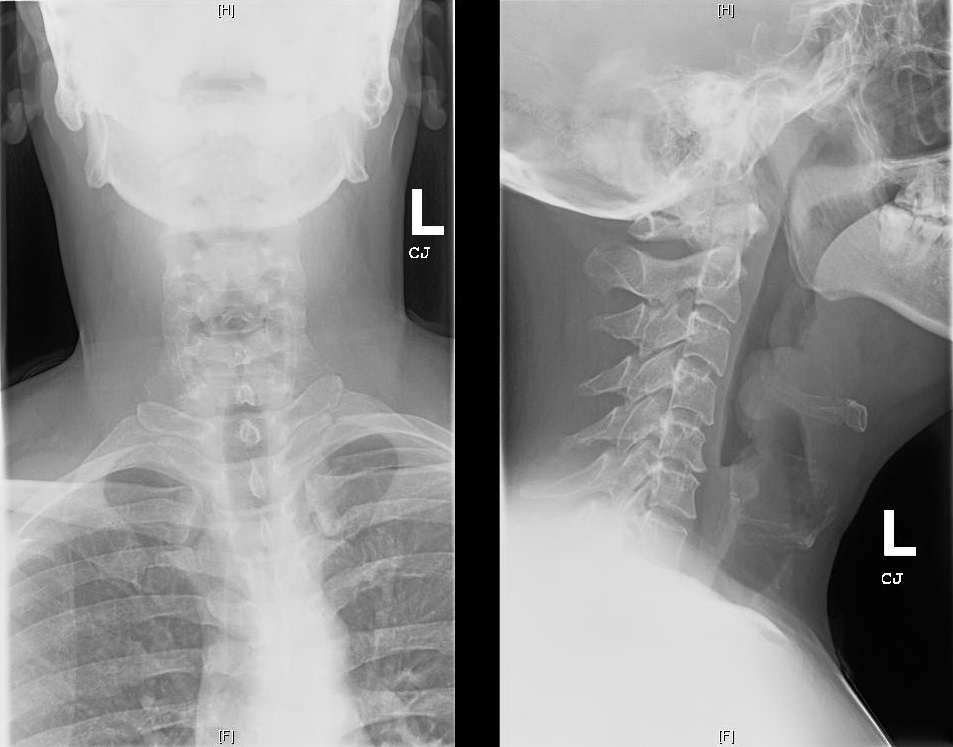
Soft Tissue Neck X-ray:

epiglottitis


THE TALK

What is an epiglottis?

  • Back wall of the vallecular space below base of tongue
  • Infectious epiglottitis = cellulitis of epiglottis
    • Progresses to involve entire supraglottic larynx (including aryepiglottic folds and arytenoids) leading to difficulty breathing

Isn’t epiglottitis a pediatric diagnosis?

  • Incidence decreased in children since haemophilus influenza vaccination
  • Incidence in adults (2006): 1.6 cases per 100,000 adults
    • Usually a/w HTN, DM, Substance abuse or immune deficiency

How do I diagnose it?

  • Fiberoptic nasal laryngoscopy = gold standard
  • Lateral neck x-ray: look for the “thumbprint” sign
  • CT neck (if diagnosis unclear)

What do I do if I suspect/diagnose it?

  • MANAGE AIRWAY- Be prepared for a cricothyrotomy!
  • Have patient sitting up in bed
  • Consider prophylactic intubation
    • Risk of laryngospasm with scope
  • Heliox (mixture of Helium and Oxygen) can be used as a temporizing measure
  • Augmentin or Ampicillin-sulbactam (Unasyn) are preferred initial antibiotics
  • Consider Vancomycin if patient is critically ill and MRSA a possible etiology
  • NSAIDS for pain control
  • +/- Steroids for symptomatic relief

This was too long and I didn’t read it- what should I know?

  • Patient with sore throat and muffled voice but no obvious signs of upper airway obstruction need further work up
  • Have your triple set up ready (direct laryngoscope, video laryngoscope and cricothyroidectomy) and have backup (ENT/Anesthesia) available for a difficult airway

REFERENCES/FURTHER READING:

  • Frantz TD, Rasgon BM, Quesenberry CP. Acute Epiglottitis in Adults: Analysis of 129 Cases. 1994;272(17):1358-1360.
  • Woods, Charles. “Epiglottitis (supraglottitis): Clinical Features and Diagnosis.” UpToDate, 23 June 2015. Web.
  • Rogers, Matt. “Epiglottitis.” Core EM, 26 Aug. 2015. Web.

Epidural Abscess

Followup Rounds
12/11/15
Article inspired by: Dr. Anna Meyendorff


THE CASE

27 y/o M p/w left upper back pain x 10 days.

  • Felt a bump on his back for the last few weeks,
  • Swelling waxed and waned
  • Took ibuprofen and has been using a heating pad for the pain.
  • Last 3 days, has developed fevers/chills, nausea, vomiting, diarrhea, and poor appetite.
  • Entire body hurts, especially his muscles
  • Cannot walk secondary to the pain and general weakness
  • Denies numbness, paresthesias, incontinence, IVDU
  • ROS- mild headache with photophobia, some difficulty swallowing and a sensation of food getting stuck in his throat

PMHx: HIV/AIDS (last CD4 6, VL 119,520), Frequent skin abscesses

PSHx: Anal wart resection

Meds: Non-compliant (prescribed: Norvir, Reyataz, Truvada, Azithromycin, Bactrim)

Allergies: Denies

SHx: Smokes 1/2ppd, occasional cocaine, denies IVDU; multiple male partners, uses condoms

Physical Exam:

  • Vitals: BP: 118/50, HR: 116, RR: 20, Temp: 102.6F, SpO2: 97%
  • General: Lying on stretcher in no distress, Cachectic, Diaphoretic, Oral thrush
  • Chest: CTA b/l, No wheezes/rales/rhonchi
  • Cardiac: Tachycardic, No murmurs/rubs/gallops
  • Abdomen: Soft, NT/ND
  • Rectal: Tone intact, No saddle anesthesia
  • Back: No midline cervical/thoracic/lumbosacral tenderness; Large tender area with local induration and calor over the left paraspinal area at mid-thoracic level
  • Neuro: AAOx3; Unable to test gait (refusing 2/2 pain); Motor 5/5 in b/l UE, 5/5 in LLE hip flexion, knee flex/ext, plantar/dorsiflexion, 3/5 RLE hip flexion, 4/5 knee flex/ext, 5/5 plantar/dorsiflexion; Sensation symmetrically intact to light touch over face, trunk, and extremities; Reflexes 3+ b/l biceps, 2+ b/l ankle jerk, toes flex b/l
  • Extremities: Warm and well-perfused x 4, Several scars on right arm (from old-appearing, small, healed abscesses), Tenderness to light touch over R anterior thigh and L triceps

Labs

  • 7.8>10.8/33<322
  • 127/4.7/89/25/19/0.9<107
  • Lactate 1.9, Alb 3.0, T. Bili 1.5, AST 198, ALT 87, Alk Phos 84
  • Coags nl, CRP 391, ESR 117

Imaging

  • CT Chest & Thoracic Spine w/o contrast: Large lobulated collection at the lateral aspect of the left paraspinal musculature concerning for a soft tissue abscess measuring 2 x 6 x 11.7 cm. Small epidural collection along the left lateral aspect of the upper thoracic spine at the T3 level. Possible fluid collection in the neuroforamina at T3/4. No osseous erosions seen. Follow-up with contrast-enhanced MRI recommended.

THE TALK

  • Two types of epidural abscesses
    • Intracranial epidural abscess
    • Spinal epidural abscess (nine times more common)
      • Most common in thoracolumbar areas (larger epidural space and more infection-prone fat tissue)

Why is an epidural abscess dangerous?

  • Can expand and compress brain/spinal cord by
    • Direct compression
    • Thrombosis/thrombophlebitis of nearby veins
    • Interruption of arterial blood supply
    • Bacterial toxins and mediators of inflammation

What does the epidural abscess contain?

  • S. aureus (63%)
  • Mycobacterium tuberculosis (more frequent cause in developing world)
  • In acute cases, frank pus
  • More commonly, granulation tissue (when present > 2 weeks)

How can bacteria get into the epidural space?

  • Hematogenously
    • Skin/soft tissue infections
    • Bacterial endocarditis
    • PNA/UTI
  • Direct extension from infected contiguous tissue
    • Vertebral osteomyelitis
    • Retropharyngeal abscess
    • Psoas abscess
  • Direct inoculation into the spinal canal
    • Epidural injections or catheters
    • Penetrating injury
    • Spinal stimulators

Who is at risk for epidural abscesses?

  • IV drug users
  • Immunocompromised (diabetes, alcoholics, HIV)
  • Recent perispinal procedures (epidural, L.P., spinal surgery)
  • Infection of adjacent structures (vertebral osteomyelitis, infected pressure sores)

Is there a classic triad I should look out for?

  • Fever (50%)
  • Back Pain
  • Neurological deficits (usually absent early on so diagnosis missed at 1st presentation)

What is the classical progression of the disease, if left untreated?

  • Back pain (focal and severe), progressing to
  • Nerve root pain (“shooting” or “electric shocks”), progressing to
  • Motor weakness, sensory changes and bladder/bowel dysfunction, progressing to
  • Paralysis (quickly becomes irreversible)

Are there any lab tests I can order to help me?

  • Leukocyte count can be elevated or normal
  • ESR is usually elevated in both epidural abscess and vertebral osteomyelitis
  • Blood cultures

Hmm, I guess not. How about imaging?

  • MRI is the preferred test (image entire spinal column because multiple skip lesions are common)
    • May require pain management so they can lie flat/still for an MRI
    • MRI is important to distinguish epidural soft tissue edema VS epidural abscess
    • Fluid-equivalent signal intensity on T2-weighted images with rim enhancement and hypointense center
  • CT with IV contrast is an acceptable alternative, if no MRI available

Okay, what do I do if the MRI suggests an epidural abscess?

  • Diagnosis
    • 2 sets of blood cultures
    • Direct needle aspiration (usually under CT guidance) of abscess fluid/pus
    • Do NOT L.P. (risk of seeding subarachnoid space leading to meningitis)
  • Treatment
    • Reduce size and ultimate elimination of inflammatory mass
      • Early surgical decompression and drainage (within 24 hours)
      • Medical (conservative) approach only if lacking risk factors (such as advanced age, bacteremia, WBC > 12,500 cell/L, diabetes, MRSA infection, in whom organism is known from aspiration and no neurological deficit)
    • Eradicate the causative organism
      • Empiric antibiotics against staphylococci, streptococci and gram-negative bacilli
        • Vancomycin (for empiric MRSA) +
        • Nafcillin OR Oxacillin (for optimal MSSA coverage as better than Vanc) +
        • Metronidazole +
        • Cefoxatime OR Ceftriaxone OR Ceftazidime (preferred if Pseudomonas considered)

REFERENCES

Meyendorff A. “Follow up Rounds: Epidural Abscess” Jacobi Medical Center. Jacobi/Montefiore Emergency Medicine Conference. Bronx. Dec 2015. Case Presentation

Sexton, Daniel J., and John H. Sampson. “Spinal Epidural Abscess.” Spinal Epidural Abscess. UpToDate, 4 Jan. 2016. Web.

Nickson, Chris. “Spinal Epidural Abscess.” Spinal Epidural Abscess. Life in the Fast Lane, Web.

PCP Pneumonia

Inspiration:
Jacobi/Montefiore Conference
Edouard Coupet, MD PGY-4

The Case:
Triage: 63 y/o M complains of SOB and cough x 3 days
Nursing Assessment: alert, making grunting noise with breathing

HPI: 63M p/w SOB. Patient notes 2 days of increasing shortness of breath. Also notes a non-productive cough. Denies any pain, but does note subjective fevers and malaise. Denies recent travel.

PMH: HTN, asthma
Meds: amlodipine, albuterol

Physical Exam

T: 101, BP: 137/68, RR: 22, HR: 118, O2: 98% on NRB
General: moderate distress
Skin: WNL
Heart: tachycardic
Lungs: coarse rales
Abd: WNL
Ext: WNL

Labs

Na: 138, Cl: 101, BUN: 32, Glucose: 117
K: 4.9, CO2: 26, Cr: 1.4

WBC: 10.2, Hgb: 8.2, Hct: 24.2, Plt: 306

Emergency Department Course

  • O2 sat improved from initial 40% on RA to 90% on non-rebreather. He was then dstarted on non-invasive positive pressure ventilation (BiPAP) 8/5 @ 100% FiO2

PCP CXR

  • CXR report: diffuse hazy airspace opacity of both lungs, may be due to CHF or pneumonia
  • The patient was started on Ceftriaxone and Azithromycin for presumed community acquired pneumonia
  • Patient states he was recently tested for HIV and was negative

Three Hours Later

  • Patient becomes increasingly hypoxic on NIPPV. Patient was intubated, but course was complicated by multiple desaturations to 70s
  • After intubation, patient was noted to be very difficult to bag. Critical care consulted
  • “Patient is now intubated and requiring a lot of ventilator support, PEEP 12, and 100% FiO2. Hemodynamically stable. Sedated now. Reason for resp failure likely pneumonia progressing to ARDS. Will cover with Vanc, Zosyn, & Azithromycin. Send urine legionella & pneumococcal antigen. CXR findings could be PCP, send PCP smears. Send HIV study if possible.”

ICU Course

  • Patient developed ARDS likely 2/2 PCP and was treated accordingly. He eventually tested positive for HIV and required ECMO and pressors in the ICU. CD4 count was very low
  • Patient was deccanulated from ECMO 9 days after presentation and extubated 3 days later. He was started on HAART and PCP prophylaxis per ID recs. He was discharged 32 days after initial presentation

PCP Pneumonia

  • Overview:
    • Caused by Pneumocystis jiroveci, a fungi, in immunocompromised patients
    • This is an AIDS defining illness
    • History usually includes shortness of breath, low fever, cough
    • Patients with PCP pneumonia can decompensate very quickly and (as in this case) go into ARDS
  • How should we diagnose it?
    • CXR might show butterfly pattern ie findings in bilateral lungs. These findings are usually interstitial
    • PCP pnuemonia increases risk of pneumothorax, which can also be seen on CXR
    • CT chest show ground glass opacities
    • High LDH (>300 U/I)
    • Send sputum cultures to help out your medicine colleagues
      • Induced sputum cultures sent to the lab get giemsa and methenamine silver stains
      • PCR is also a mode of diagnosis
    • How should it be treated?
      • Emperic therapy immediately. PCP pneumonia has a very high morbidity and mortality
      • First line treatment is TMP-SMX either PO or IV (use IV for more severe cases)
        • Trimethoprim 20mg/kg/day and sulphamethoxazole 150mcg/kg/day in 4 divided doses for 21 days
      • Alternatives: primaquine + clindamycin, atovaquone, dapsone + trimethoprim
      • If patient is HIV positive, steroids will help
    • Propylaxis: If CD4 < 200 cells/mm or if HIV positive with history of oropharyngeal candidiasis

References
Coupet E. “M&M Conference: PCP Pneumonia.” Jacobi Medical Center. Jacobi/Montefiore Emergency Medicine Conference. Bronx. Dec 2015. Lecture

Nickson, Chris. “Pneumocystis Jiroveci Pneumonia.” Life in the Fast Lane. Web. 26 Jan. 2016. http://lifeinthefastlane.com/ccc/pneumocystis-jiroveci-pneumonia/

Malaria

Conference
11/4/2015
Presenter: Edouard Coupet, MD

THE CASE
CC: “Joint pain”
HPI:
  • 23 yo F sent to ED by PMD for “joint pain”
  • Reports polyarthralgias x 1 day
  • Denies CP or SOB
  • Subjective fevers
  • Visited Nigeria 4 weeks prior
  • Did not take malaria ppx
PMH/PSH: HbSS
Medications: Denies
Allergies: NKMA
Physical Exam
Vitals: T 97.1, HR 102, BP 143/73, RR 16, O2 97% on RA
General: NAD
Skin: Jaundiced
HEENT: Scleral icterus
CVS: Tachycardic, regular rhythm, no murmurs
Pulm: CTAB
Extremities: No erythema, no visible e/o trauma, FROM
Studies: 
CBC: Hgb 4.1, Hct 11.3, WBC 20.4, Plt 98, Retic 11.2
Chem: Na 137, K 3.3, CO2 19, Gluc 127, AG 22
LFTs: T. bili 40.1, D. bili 22.8, AST 226, ALT 91, Alk P 247

MALARIA
Background:
  • Most common cause of hemolytic anemia worldwide
  • Most significant disease acquired from travel to tropics
  • P. falciparum most deadly parasite; most common form in Africa, Haiti, New Guinea
  • P. vivax more common in Central America, Indian subcontinent
  • Incubation period = 8-25 days (generally)
  • Mortality = 10-50% if untreated
  • Clinical signs of infection occur during the erythrocytic phase
    • Cyclical symptoms = erythrocytes lyse due to intracellular replication → merozoites released → new erythrocytes infected
  • Parasitized cells lose flexibility → obstruct microcirculation → tissue anoxia of lungs/kidneys/brain
  • Parasite may not be visualized on smear due to sequestration
How do I make the diagnosis?
  • Symptoms
    • Periodic fevers
    • Malaise
    • HA
    • CP
    • Cough
    • Abd pain
    • Arthralgia
    • Diarrhea
  • Paroxysms may not be present in patient’s who received chemoprophylaxis
  • Splenomegaly, abdominal tenderness
  • Anemia, jaundice
  • Coma, altered mental status
  • Blood smear
    • First smear positive in >90% of cases
    • If initial negative, must be repeated BID x 2-3 days for proper exclusion of malaria
    • Determines degree of parasitemia and type (i.e. P. falciparum)
  • Additional lab findings
    • Normocytic anemia
    • Thrombocytopenia
    • ↑ ESR
    • ↑ LDH
    • LFT abnormalities
    • ↑ Cr
    • Hyponatremia
    • Hypoglycemia
    • False positive VDRL
What makes malaria deadly?
  • Hemolysis can lead to severe anemia
  • Sequestration → splenomegaly → splenic rupture
  • Immune mediated glomerulonephritis
  • Cerebral edema (mortality rate >20%)
    • LP: ↑ opening pressure, ↑ protein, mild pleocytosis
  • Noncardiogenic pulmonary edema
  • Renal failure (ATN)
  • Hypoglycemia
  • Adherence of infected cells to endothelium → tissue hypoxia
What is “uncomplicated” P. falciparum and how is it managed in the ED?
  • Uncomplicated:
    • No e/o organ dysfunction
    • Parasitemia <5%
    • Able to tolerate PO
  • Hospitalize:
    • Severe clinical manifestations in non-immune host for P. falciparum or P. knowlesi 
  • Report to state health department
  • For non-pregnant patients (3 day course)
    • Artemether + lumefantrine
    • Artesunate + amodiaquine
    • Artesunate + mefloquine
    • Dihydroartemisinin + piperaquine
    • Artesunate + sulfadoxine–pyrimethamine (SP)
  • For pregnant (1st trimester)
    • Quinine + clindamycin x 7 days
  • Additional considerations
    • Avoid artesunate + SP in HIV/AIDS patients taking co-trimoxazole
    • Avoid artesunate + amodiaquine in HIV/AIDS patients taking efavirenz or zidovudine
And if it’s severe?
  • Severe P. falciparum malaria:
    • GCS < 11
    • Generalized weakness
    • >2 convulsions within 24 hours
    • Acidosis
    • Shock
    • Pulmonary edema
    • Cr > 3 mg/dL or BUN >20
    • T. bili >50 + parasite count >100,000
    • Significant hemorrhage
    • Hgb <7 g/dL, Hct <20%
    • Glucose <40 mg/dL
    • Hyperparasitemia >10%
  • Do not delay treatment in the unstable patient if strong suspicion for malaria as initial smear may be falsely negative
  • Treatment (IV for ≥24 hours then 3 days PO course)
    • Artesunate (IV)
      • Clears malaria faster than quinine
      • Distributed only through CDC
    • Quinidine (IV) also appropriate choice; more available in US
What do I do for the associated complications?
  • AMS
    • Due to cerebral sequestration of infected erythrocytes
    • Requires LP to rule out meningitis as cause
    • Seizures commonly reported in children and to be treated with benzodiazepines
    • Steroids of no benefit
  • Anemia
    • Transfuse at Hgb <4 g/dL or <6 g/dL if AMS, acidosis, shock, or parasitemia >20%
    • There is no evidence to support exchange transfusions as an adjunct and is not recommended by CDC or WHO
  • Respiratory distress
    • Patient may develop noncardiogenic pulmonary edema (similar to ARDS)
    • Supplemental O2 to be administered
    • May require mechanical ventilation

REFERENCES

Bremen, Joel. “Clinical manifestations of malaria.” Up To Date. http://www.uptodate.com, 5 Nov. 2015. Web. 30 Nov. 2015. http://www.uptodate.com/contents/clinical-manifestations-of-malaria

Kyriacou DN, Spira AM, Talan DA, Mabey DC. Emergency department presentation and misdiagnosis of imported falciparum malaria. Ann Emerg Med. 1996;27(6):696-9.

Taylor, Terrie. “Treatment of severe falciparum malaria.” Up To Date. http://www.uptodate.com, 29 Oct. 2015. Web. 30 Nov. 2015. http://www.uptodate.com/contents/treatment-of-severe-falciparum-malaria

Tintinalli, Judith E., and J. Stephan. Stapczynski. Tintinalli’s Emergency Medicine: A Comprehensive Study Guide. 7th ed. New York: McGraw-Hill, 2011.

World Health Organization. Guidelines for the treatment of malaria, 3rd ed, WHO, Geneva 2015. http://www.who.int/malaria/publications/atoz/9789241549127/en/